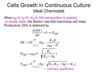
Download

1 / 24
240 likes | 368 Vues
Plutonium interactions with cells in culture. Tatjana Paunesku , Baikuntha Aryal , Chuan He, Drew Gorman-Lewis, Stefan Vog , Barry Lai, Lynda Soderholm , Mark Jensen, and Gayle Woloschak Department of Radiation Oncology, Northwestern University, Chicago, IL USA

E N D
Plutonium interactions with cells in culture Tatjana Paunesku, BaikunthaAryal, Chuan He, Drew Gorman-Lewis, Stefan Vog, Barry Lai, Lynda Soderholm, Mark Jensen, and Gayle Woloschak Department of Radiation Oncology, Northwestern University, Chicago, IL USA Department of Chemistry, University of Chicago, Chicago, IL, USA Chemical Science and Engineering Division, Argonne National Laboratory, Argonne, IL USA X-ray Sciences Division, Advanced Photon Source, Argonne National Laboratory, Argonne, IL USA The First International Conference on Radiation and Dosimetry in Various Fields of Research (RAD 2012), Niš, Serbia, April 25-27, 2012.
Biological effects of plutonium (Pu) exposure • Regardless of the entry route, Pu deposition in the organism can last for decades. • Pu effects on cells and organisms depend not only on alpha particle emission, but also on behavior of Pu as a heavy metal. • Biochemical interactions of Pu are of great interest for investigation and interpretation of Pu effects on living cells and organisms.
Interaction of Pu with cells in culture Short term interaction of Pu with cells was studied in vitro. • Cellular entry of Pu, and differentiation of different chemical states of Pu was investigated using synchrotron X-rays (Gorman-Lewis et al., 2011; Jensen et al., 2011; Jensen et al., 2012) • Transferrin bound Pu is taken up by the cells in the form of tetravalent Pu. (Jensen et al 2011) • Intracellular Pu(IV) interacting proteins were studied by metaloproteomicsapproaches . (Aryal et al., 2011)
(1) Study of plutonium-cells interactions using X-ray microscopy and spectroscopy
X-ray microscopy and spectroscopy for Pu detection and chemical speciation • X-ray fluorescence microscopy (XFM) with hard X-rays can be used to map and quantify elemental make-up of cells and tissue samples. • Most native biological elements and trace elements are detected by their K alpha characteristic fluorescence. • High Z elements including actinides such as plutonium (Pu) can be detected by hard X-ray XFM by their L lines characteristic fluorescence. • Valence state of Pu can also be investigated by microspectroscopictechiquemicrobeam X-ray absorption near edge structure (µ-XANES).
Advanced Photon Source: Argonne National Laboratory Elemental distribution in cells is mapped by detecting element specific K(or L lines) X-ray fluorescence at one of the beamlines of the X-ray Operations and Research Collaborative Access Team (XOR-CAT) at the Advanced Photon Source of Argonne National Laboratory.
XFM schematic • Example APS 2-ID-D: • energy range E = 5 – 30 keV • spatial resolution = 150 nm • focussed flux 2·109 phot/s (can trade flux for resolution) • stepscan sample through focused X-ray beam • record full XRF spectrum at each scan point • compare specimen counts/spectra to calibration curve, to quantify to area density Courtesy of S. Vogt, APS, Argonne National Laboratory, USA
Intensity Energy X-ray induced X-ray fluorescence – a brief reminder Emission of Auger e- - dominating low Z photo-electric absorption of incident hard X-ray emission of photo-electron X-ray fluoresence - dominating high Z Detect XRF using energy dispersive detector • Energy of X-ray fluorescence photons is characteristic for each element • XRF is quantitative, i.e., number of XRF photons is directly related to quantity of element • Photo-electric absorption crossection straightforward to calculate (monochomatic incident beam)
Pu is localized in cytoplasm—overlap with Fe? • PC12 cells were treated with 242Pu. Two individual cell images show that: • (1) Pu (red) and Fe (green) co-localize (yellow) --- initial hypothesis that Fe and Pu enter cell by the same mechanism • (2) Pu (red) and Zn in upper cell (blue) or P (green) in lower cell do not co-localize --- since P and Zn are the highest in the nucleus, Pu apparently remains in the cytoplasm Jensen et al., 2011, Nature Chemistry
Pu X-ray absorption spectroscopy—only tetravalent Pu is inside cells The intracellular oxidation state of plutonium was measured directly using microprobe X-ray absorption near edge spectroscopy. Despite the generally reducing conditions found inside living cells and regardless of the initial oxidation state or chemical form of plutonium presented to rat adrenal gland cells, the X-ray absorption spectra of the intracellular deposits were always consistent with tetravalent Pu. Gorman-Lewis et al., 2011 Inorganic Chemistry Jensen et al., 2012. AnalyticaChimicaActa
Pu X-ray absorption spectroscopy—only tetravalent Pu is inside cells Average plutonium content of individual cells incubated for 3 hours in complete media with serum containing 100 μMPu added as Pu(III), Pu(IV), or Pu(VI). The corresponding average total Pu contents are Pu(III), 1.7 ± 0.2 fg per cell; Pu(IV), 15.8 ± 3.2 fg per cell; and Pu(VI), 2.1 ± 0.5 fg per cell. Uncertainties are given as the standard error of the mean for measurement of n cells. Gorman-Lewis et al., 2011 Inorganic Chemistry
(2) Study of plutonium and transferrin interaction by small angle x-ray scattering
X-ray small angle scattering (SAX) • SAX allows evaluation of proteins in solution • Rough protein structures reconstructed from SAX data can be compared with existing protein crystal structures • Testing hypothesis that Pu enters cells using same mechanism as Fe—SAX of transferrin molecules with different quantity and location of Pu and Fe
Structural models of transferrins with different quantities and locations of Fe and Pu derived from SAXS: --PuCFeNTf (green) adopts a conformation most similar to the native Fe2Tf (yellow) (a)Orthogonal views of the three-dimensional molecular envelopes of metal–Tf structures reconstructed from the SAXS. (b) Docking wireframe representations of the Tf structures superimposed onto each other Jensen et al., 2011, Nature Chemistry
X-ray studies of cellular Pu: Conclusions • Pu (IV) enters cells most abundantly • Pu (IV) can imitate Fe and enter cells using transferrin uptake route • Once inside cells Pu is released from tranferrin and can interact with intracellular proteins
Discovery of intracellular Pu(IV) interacting proteins: approach A mixture of cellular proteins was loaded onto a Pu(IV) NTA (plutonium bound to nitrilotriacetic acid), after appropriate washes and elution steps many cellular proteins other than transferrin were found to bind to Pu. Most Pu binding proteins detected by 2DE are metalloproteins binding Ca2+, Zn2+ or other divalent ions. Computational MetaCore™ v.6.2 analysis shows that majority of the cellular proteins interacting with Pu have pro-neoplastic roles (same is true for transferin receptors which can bind Pu loaded transferrin). Aryal et al., 2011, J. Proteome Res
Proteins interacting with Pu(IV)-NTA 3 pI 10 Mwt Silver stained 2D PAGE of proteins isolated from rat pheochromocytoma PC12 cells previously panned on Pu(IV)-NTA IMAC. Proteins particularly discussed here are labeled as spots 1 to 7. Aryal et al., 2011, J. Proteome Res
Plutonium binding proteins identified by LC-MS/MS analysis Aryal et al., 2011, J. Proteome Res
MetaCore™ v.6.2 analysis was used to construct a "shortest distance pathway" connecting the proteins identified to be involved in Pu binding, directly or indirectly (via transferrin) (box). Every arrow represents a curated relationship between proteins connected; red arrows are inhibitory interactions, green arrows activations and gray arrows indicate unspecified (or more complex) relationships. One "upstream" and one "downstream" relationship from GRP78 are highlighted by light blue as examples. Aryal et al., 2011, J. Proteome Res
MetaCore™ v.6.2 analysis : GO Processes for a "shortest distance" network including Pu binding proteins: GRP78, actin gamma1, NDPK B, pyruvatekinase and galectin 1. Aryal et al., 2011, J. Proteome Res
MetaCore™ v.6.2 analysis :Diseases associated with the collection of proteins involved in the "shortest distance" pathway Aryal et al., in press, J. Proteome Res
Pumetaloproteomics: Conclusions • Pu (IV) interacting cellular proteins have pro-neoplastic roles • Puloaded proteins probably do not fulfill their cellular roles—competition with divalent metals (Ca2+ , Mg 2+, Zn 2+,…) as well as Fe • Possible feedback loop where Pu caused neoplastic transformation leads to an increase of Pu interacting proteins and/or increased Pu load?
Thank you! Lab picnic 2010