Download
1 / 82
840 likes | 1.28k Vues
Lisa Randall, RN, MSN, ACNS-BC RNSG 2432. Cerebrovascular Accident “Brain Attack”. Objectives. Define cerebrovascular accident and associated terminology Discuss related pathophysiology and presentation of various types of stroke

E N D
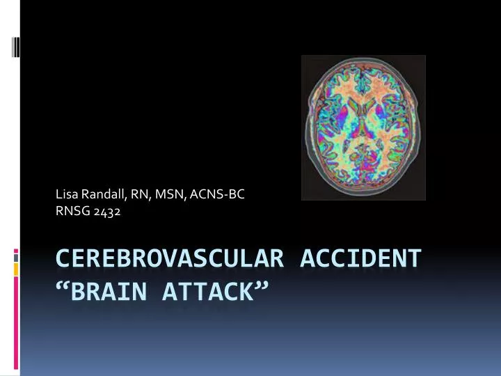
Lisa Randall, RN, MSN, ACNS-BC RNSG 2432 Cerebrovascular Accident“Brain Attack”
Objectives • Define cerebrovascular accident and associated terminology • Discuss related pathophysiology and presentation of various types of stroke • Discuss etiology, risk factors, diagnostics, management, and outcomes of stroke • Review case studies and nursing diagnoses, interventions, and goals
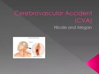
Definition • Stroke or “brain attack” is an acute CNS injury that results in neurologic S/S brought on by a reduction or absence of perfusion to a territory of the brain. The disruption in flow is from either an occlusion (ischemic) or rupture (hemorrhagic) of the blood vessel.
Incidence & Prevalence • Third leading cause of death in the USA • 750,000+ people/year • 175,000 die within one year (25%) • Leading cause of long-term disabilities • 5.5 million survivors (USA) • 15 to 30 % live with permanent disability
Definitions • Cerebrovascular Accident • Ischemic Stroke • Thrombotic • Embolic • Lacunar infarct • TIA • Hemorrhagic Stroke • ICH • SAH
Stroke: Emergency Care • http://youtu.be/-d8__FkW-nU
Thrombotic Stroke • Occlusion of large cerebral vessel • Older population • Sleeping/resting • Rapid event, but slow progression (usually reach max deficit in 3 days)
Embolic Stroke • Embolus becomes lodged in vessel and causes occlusion • Bifurcations are most common site • Sudden onset with immediate deficits • Embolysis • Hemorrhagic Transformation
Lacunar Strokes - 20% of all stokes • Minor deficits • Paralysis and sensory loss • Lacune • Small, deep penetrating arteries • High incidence: • Chronic hypertension • Elderly • DIC
Transient Ischemic Attack • Warning sign for stroke • Brief localized ischemia • Common manifestations: • Contralateral numbness/ weakness of hand, forearm, corner of mouth • Aphasia • Visual disturbances- blurring • Deficits last less than 24 hours (usually less than 1 or 2 hrs) • Can occur due to: • Inflammatory artery disorders • Sickle cell anemia • Atherosclerotic changes
Hemorrhagic Stroke Definitions • Intracerebral hemorrhage • Intracranial hemorrhage • Parenchymal hemorrhage • Intraparenchymal hematoma • Contusion • Subarachnoid hemorrhage
Hemorrhagic Stroke • Rupture of vessel • Sudden • Active • Fatal • HTN • Trauma • Varied manifestations
Hemorrhagic Stroke • Intracerebral Hemorrhage • Subarachnoid Hemorrhage
PathophysiologyHemorrhagic Stroke • Changes in vasculature • Tear or rupture • Hemorrhage • Decreased perfusion • Clotting • Edema • Increased intracranial pressure • Cortical irritation
Legs Mom: Bowel/bladder Reasoning/judgment Long term memory Voluntary Motor Sensations Pain & Touch Taste Arms Head Vision & visual memory Hearing/association & Smell & taste Short term Memory Balance, Coordination of each muscle group CN 5,6,7,8 P,R, B/P CN 9,10,11,12 Tracks cross over Coordinate movement, HR,B/P
Vessels of the Brain Right Side
PhysiologyNormal Cerebral Blood Flow • Oxygen • Glucose • 20% of Cardiac Output / oxygen • Arterial supply to the brain: • Internal carotid (anteriorly) • Vertebral arteries (posteriorly) • Venous drainage • 2 sets of veins - venous plexuses • Dural sinuses to internal jugular veins • Sagittal sinus to vertebral veins • No valves, depend on gravity and venous pressure gradient for flow
Risk Factors NON-MODIFIABLE MODIFIABLE • Age • 2/3 over 65 • Gender • M=F • Female>fatality • Race • AA > hispanics, NA • Asians > hem • Heredity • Family history • Previous TIA/CVA • Hypertension • Diabetes mellitus • Heart disease • A-fib • Asymptomatic carotid stenosis • Hyperlipidemia • Obesity • Oral contraceptive use • Heavy alcohol use • Physical inactivity • Sickle cell disease • Smoking • Procedure precautions
EtiologyIschemic Stroke Embolism Prothrombotic states • Atrial fib • Sinoatrial D/O • Recent MI • Endocarditis • Cardiac tumors • Valvular D/O • Patent foramen ovale • Carotid/basilar artery stenosis • Atherosclerotic lesions • Vasculitis • Hemostatic regulatory protein abnormalities • Antiphospholipid antibodies • Hep cofactor II
Etiology Hemorrhagic Stroke • Chronic HTN** • Cerebral AmyloidAngiopathy* • Anticoagulation* • AVM • Ruptured aneurysm (usually subarachnoid) • Tumor • Sympathomimetics • Infection • Trauma • Transformation of ischemic stroke • Physical exertion, Pregnancy • Post-operative
Aneurysm • Localized dilation of arterial lumen • Degenerative vascular disease • Bifurcations of circle of Willis • 85% anterior • 15% posterior
AneurysmSubarachnoid Hemorrhage • SAH • Mortality 70% • 97% HA • Nuchal rigidity • Fever • Photophobia • Lethargy • Nausea • Vomiting
Aneurysm/SAH • Complications • HCP • Vasospasm • Triple H Therapy • HTN • Hemodilution • Hypervolemia • Surgical treatment • Clip • Coil • INR
Nursing Management • Assessment • Monitoring • BP • TCDs • CBC • Preventing complications • Bowel program • DVT prophylaxis • Siezure prophylaxis • Psychological support • Discharge planning
Arteriovenous malformations • AVM • Tangled mass of arteries and veins • Seizure or ICH
Treatment AVM • Endovascular • Neurosurgery • Radiosurgery
Presentation • Sudden onset • Focal neurological deficit • Progresses over minutes to hours • HA, N/V, <<LOC, HTN • Depends on location
Stroke Symptoms include: • SUDDEN numbness or weakness of face, arm or leg • SUDDEN confusion, trouble speaking or understanding. • SUDDEN trouble with vison. • SUDDEN trouble walking, dizziness, loss of balance or coordination. • SUDDEN severe HA.
Manifestationsby Vessel • Vertebral Artery • Pain in face, nose, or eye • Numbness and weakness of face (involved side) • Gait disturbances • Dysphagia • Dysarthria (motor speech)
Manifestationsby Vessel • Internal carotid artery • Contralateral paralysis (arm, leg, face) • Contralateral sensory deficits • Aphasia (dominant hemisphere involvement) • Apraxia (motor task), • Agnosia (obj. recognition), • Unilateral neglect (non-dominant hemisphere involvement) • Homonymous hemianopia
Manifestations & Complications by Body System • Neurological • Hyperthermia • Neglect syndrome • Seizures • Agnosias (familiar obj) • Communication deficits • Aphasia (expressive, receptive, global) • Agraphia • Visual deficits • Homonymous hemianopia • Diplopia • Decreased acuity • Decreased blink reflex
Manifestations & Complications by Body System • Neurological (cont.) • Cognitive changes • Memory loss • Short attention span • Poor judgment • Disorientation • Poor problem-solving ability • Behavioral changes • Emotional lability • Loss of inhibitions • Fear • Hostility
Manifestations & Complications by Body System • Musculoskeletal • Hemiplegia or hemiparesis • Contractures • Bony ankylosis • Disuse atrophy • Dysarthria - word formation • Dysphagia – swallow • Apraxia – complex movements • Flaccidity/spasticity • GU • Incontinence • Frequency • Urgency • Urinary retention • Renal calculi
Manifestations & Complications by Body System • Integument • Pressure ulcers • Respiratory • Respiratory center damage • Airway obstruction • Decreased cough ability • GI • Dysphagia • Constipation • Stool impaction
Initial Stroke Assessment/Interventions • Neurological assessment & NIH assessment • Call “Stroke Alert” Code • Ensure patient airway • VS • IV access • Maintain BP within parameters • Position head midline • HOB 30 (if no shock/injury) • CT, blood work, data collection/NIH Stroke Scale • Anticipate thrombolytic therapy for ischemic stroke
NIH Stroke Scale Score • Standardized method • measures degree of stroke r/t impairment and change in a patient over time. • Helps determine if degree of disability merits treatment with tPA. • As of 2008 stroke patients scoring greater than 4 points can be treated with tPA. • Standardized research tool to compare efficacy stroke treatments and rehabilitation interventions. • Measures several aspects of brain function, including consciousness, vision, sensation, movement, speech, and language not measured by Glasgow coma scale. • Current NIH Stroke Score guidelines for measuring stroke severity: Points are given for each impairment. • 0= no stroke • 1-4= minor stroke • 5-15= moderate stroke • 15-20= moderate/severe stroke • 21-42= severe stroke • A maximal score of 42 represents the most severe and devastating stroke.
Question • The neurologic functions that are affected by a stroke are primarily related to • A. the amount of tissue area involved. • B. the rapidity of the onset of symptoms. • C. the brain area perfused by the affected artery. • D. the presence or absence of collateral circulation.
Question • A patient is admitted to the hospital with a left hemiplegia. To determine the size and location and to ascertain whether a stroke is ischemic or hemorrhagic, the nurse anticipated that the health care provider will request a • A. CT scan. • B. lumbar puncture. • C. cerebral angiogram. • D. PET scan.
Diagnostics Tests for the Emergent Evaluation of the Patient with Acute Ischemic Stroke • CT head (-) • Electrocardiogram • Chest x-ray • Hematologic studies (complete blood count, platelet count, prothrombin time, partial thromboplastin time) • Serum electrolytes • Blood glucose • Renal and hepatic chemical analyses • National Institute of Health Scale (NIHSS) score
Diagnostics Ischemic Stroke Hemorrhagic Stoke
BP MAP CPP Factor VII, Vit K, FFP ICP HOB Sedation Osmotherapy Hyperventilation Paralytics Fluid management euvolemia Seizure prophylaxis Keppra Dilantin Sedation Body temperature PT/OT/ST DVT prophylaxis Medical Management
Treatment Ischemic Hemorrhagic • Medical management • TpA • Endovascular • Carotid endarectomy • Merci clot removal • http://youtu.be/P2TNz-TniIA • Medical management • Decompression • Craniotomy • Craniectomy PT/OT/ST REHABILITATION
Medications • Anti-coagulants – A fib & TIA • Antithrombotics • Calcium channel blockers – Nimotop (nimodipine) • Corticosteroids ??? • Diuretics – Mannitol, Lasix (Furosemide) • Anticonvulsants – Dilantin (phenytoin) or Cerebyx (Fosphenytoin Sodium Injection) • Thrombolytics - tPA (recombinant tissue plasminogen activator)