Download
1 / 10
100 likes | 104 Vues
Untreated MM1 15 m M GRN163L 15 m M. A. B. Supplemental Figure S1. Effects of imetelstat on CFU-MK colony formation.

E N D
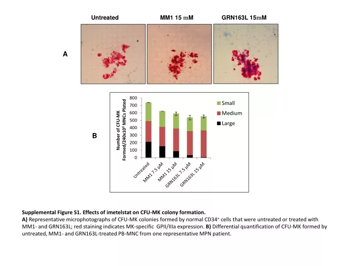
Untreated MM1 15 mM GRN163L 15mM A B Supplemental Figure S1. Effects of imetelstat on CFU-MK colony formation. A) Representative microphotographs of CFU-MK colonies formed by normal CD34+ cells that were untreated or treated with MM1- and GRN163L; red staining indicates MK-specific GPII/IIIa expression. B) Differential quantification of CFU-MK formed by untreated, MM1- and GRN163L-treated PB-MNC from one representative MPN patient.
Untreated MM1 15 mM GRN163L 15 mM 4.8% 24.7% 2.1% A 28.1% 12.3% 38.6% B CD41 CD42b Supplemental Figure S2. Effects of imetelstat on the normal MK phenotype. Representative flow cytometric analyses of MK cultures that were untreated, treated with the 15 mM control oligonucleotide MM1 or treated with 15 mM imetelstat (GRN163L) for 14 days. Oval gates indicate indicate CD34+/CD41+ MK precursors on the plots in A) and CD41+/CD42b+/high mature MK on the plots in B). CD41 CD34
B A ns p = 0.0064 ns p = 0.0152 ns ns Supplemental Figure S3. Quantitative evaluation of the effects of imetelstat on MK phenotype. Percentage of CD34+/CD41+ MK precursors (A) and mature CD41+/CD42b+ mature MK (B) detected in MK cultures that were untreated, treated with 7.5 and 15 mM of either MM1 or imetelstat (GRN163L). The columns represent the mean ± SD of three independent experiments initiated with CD34+ cells from three different donors. ns, not statistically significant.
A B Day 7 ns ns Day 14 Untreated MM1 15 mM GRN163L 15 mM CD42+/high CD42-/low Supplemental Figure S4. Effects of imetelstat on normal MK maturation. A) Representative flow cytometric analyses of CD41/CD42b expression in MK cultures that were cultured in the absence of drugs for 7 days (upper panel) then allowed to mature in the absence (untreated) and presence of 15 mM of either MM1 or imetelstat (GRN163L) for 7 additional days (lower panels). Oval gates indicate CD41+/CD42bpositive/low (green oval gate) and CD41+/CD42bpositive/high (red oval gate) mature MK. B) Frequency of CD41+/CD42b+ MK precursors quantified in MK cultures that were untreated, treated with 7.5 and 15 mM of either MM1 or imetelstat (GRN163L) as indicated in A. The columns represent the mean ± SD of three independent experiments initiated with CD34+ cells from three different donors. ns, not statistically significant. CD42b CD42b CD41 CD41
A B C ns p = 2.98E-05 E D GRN163L Untreated MM1 hTERT GAPDH Supplemental Figure S5. Evaluation of hTERT expression in normal and malignant MK. A) mRNAs extracted from normal MK cultures at various time points during differentiation and maturation (days 3, 7, 9 and 11) were reverse transcribed then amplified by quantitative real-time PCR using human TERT primers. B) mRNAs extracted from untreated normal MK cultures and from cultures treated with GRN163L for 7 days were reverse transcribed then amplified by quantitative real-time PCR using human TERT primers. C) mRNAs extracted from untreated MPN MNC cultures and from cultures treated with GRN163L for 7 days were reverse transcribed then amplified by quantitative real-time PCR using human TERT primers (n = 4 MPN patients). The y axis indicates number of PCR cycles required for hTERT mRNA amplification normalized to GAPDH. D) Western blot analyses of protein lysates isolated from JAK2V617F+ HEL MK cell line untreated or treated with MM1 and GRN163L for 72 hours. After blotting, the membranes were incubated with anti-hTERT antibodies and anti-GAPDH antibodies as control for protein loading. E) TA measured in JAK2V617F+ HEL MK cell line treated as in D). The y axis indicates the number of PCR cycles (Ct) required for the amplification of telomere repeats. A low Ct value indicates a cell extract with high TA whereas a high Ct value indicates a cell extract with low TA.
B MFI MFI MFI A Untreated MM1 15 mM GRN163L 15mM C D Untreated MM1 15 mM GRN163L 15 mM p=0.0348 p=0.0388 CD41 Supplemental Figure S6. Effects of imetelstat on pro-platelets extension and platelet production by normal human MK. A) Microscopic visualization of pro-platelets generated by MK in untreated cultures and in cultures treated with 15 mM of MM1 or GRN163L. Representative microphotographs of MK-specific GPIIb/IIIa-labeled cultures show the presence of MK bearing long pro-platelets with nascent platelet buds at their ends (arrows in a, b and c) and GPIIb/IIIa positive platelet-sized particles (arrow heads in a, b and c). B) Representative phenotypical analyses of culture-derived platelets. CD41 expression (x axis) and thiazole orange (y axis) labeling of culture-generated platelets was determined after analytical gates were set up using freshly isolated human PB platelets that were used as control; arrow indicates the measured mean fluorescence intensity (MFI) quantified in D). C) Quantification of platelets generated by untreated MK and by MK treated with 7.5 and 5 mM of MM1 or GRN163L. D) MFI ± SD observed in three independent analyses in the same culture conditions indicated in C). The columns in C) and D) represent the percentage and MFI, respectively in CD41+/TO+ platelet-sized cells ± SD detected in MK cultures derived from CD34+ cells derived from three different healthy donors analyzed independently. TO
A C * * B ns Untreated MM1 GRN163L PT5 PT141 Supplemental Figure S7. Effects of imetelstat on MPN megakaryopoiesis. Percentage of CD34+/CD41+ MK precursors (A) and CD34-/CD41+ more mature MK (B) detected in MNC cultures derived from healthy controls (HC) and MPN patients that were untreated or treated with 15 mM of either MM1 or imetelstat (GRN163L); n = 6 HC, n = 12 MPN; ns, not statistically significant. C) Representative flow cytometric analyses of CD41 (x axis) and CD42b (y axis) expression after exposure to 15 mM MM1 or GRN163L of PB MNCs cultures derived from two MPN patients (PT5 and PT141). Upper-right quadrant comprises less mature CD41+/CD42blow MK and fully mature CD41+/CD42b+/high MK (red oval gate). CD42-APC CD41-FITC
Un-labeled Untreated MM1 GRN163L PNA-FITC DNA Supplemental Figure 8. Evaluation of of imetelstat on-target binding. Untreated and MM1- or GRN163L-treated JAKV617F-positive HEL cells were labeled with FITC-conjugated telomere-specific PNA (peptide nucleic acid) and analyzed by flow cytometry. Unlabeled cells were utilized as negative control for fluorescence intensity (y axis) which was acquired in parallel with PI staining of DNA (x axis). Changes in FITC intensity is illustrated by events acquired in gate P4. Basal fluorescence resulted from PNA binding of chromosomal telomere sequences is shown in untreated and MM1-treated cultures. Increased FITC fluorescence resulted from binding of PNA to chromosomal sequences and imetelstat is shown in GRN163L-treated cultures.
A B ns ns Supplemental Figure 9. Effects of imetelstat on megakaryopoiesis in different MPN types and genotypes. Comparison between MNC cultures from MPN patients with different types of disease (i.e. MF vs. ET) and genotypes for their ability to generate CFU-MK (A) and MK cells in liquid culture (B) in the presence of imetelstat as detailed in Supplemental Table S1. ns, not statistically significant.
Imetelstat effects on MPN MK: • Inhibition of CFU-MK • Delayed MK maturation • Reduction of fibrogenicGF secretion MK Precursors (CD34+/CD41+/CD42b-) Hematopoietic Stem/Progenitor Cells Immature MK (CD34-/CD41+/CD42b-) Mature MK (CD34-/CD41+/CD42b+) Platelets • Imetelstat effects on normal MK: • Delayed maturation (reversible) Supplemental Figure S10. Proposed model for imetelstat’s mechanims of action on normal and MPN megakaryopoiesis.