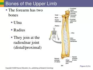
Download
1 / 27
300 likes | 683 Vues
Bones of the Lower Limb. The Dance Hal l by Vincent van Gogh ,1888. Kaan Yücel M.D., Ph.D. 16.January.2014 Thursday. 2 functional components: Pelvic girdle & bones of the free lower limb Body weight is transferred Vertebral column ( Sacroiliac joints)

E N D
Bones of theLower Limb The Dance Hall byVincent van Gogh ,1888 Kaan Yücel M.D., Ph.D • 16.January.2014 Thursday
2 functional components: Pelvic girdle & bones of the free lower limb Body weight is transferred Vertebral column (Sacroiliac joints) Pelvic girdle (Hip joints) Femurs (L. femora) Skeleton of thelowerlimb (inferiorappendicularskeleton)
FEMUR longest and heaviest bone Transmits body weight from the hip bone to the tibia. Superior / Proximal end Shaft (Body) Inferior/ Distal end
Proximalend of femur Superior (proximal) end of the femur • Head • Neck • 2 trochanters • Greater& Lesser • intertrochantericline • intertrochantericcrest • quadratetubercle • fovea capitisforlig.teres
Shaft of femur Superior (proximal) end of the femur Gluteal tuberosity Linea aspera Medial and lateral lips of lineaaspera Medial and lateral supracondylar lines Pectineal line
Superior (proximal) end of the femur Distalend of femur Adductor tubercle Intercondylar fossa Medial and lateral condyles Medial and lateral epicondyles Medial and lateral femoral condyles Patellar surface
TIBIA Located on the anteromedial side of the leg Second largest bone in the body Flaresoutward at both ends to provide an increased area forarticulation and weight transfer.
Proximalend of tibia widensto form medial& lateralcondyles (1,2) flatsuperiorarticularsurfacetibialplateau (3) articularsurfacesseparatedby intercondylareminence (4) formedby 2 intercondylartubercles medialandlateral (5,6) flankedbyrelativelyrough anteriorandposteriorintercondylarareas (7,8) • 1 • 4 • 5 • 6 2 Anterolateralview of lefttibia
medialmalleolus Interosseous membrane unites the two leg bones. Inferiorly, the sharp border is replaced by fibular notch. Distalend of tibia
PATELLA (Kneecap) • Largestsesamoid bone (a bone formed within the tendon of a muscle) in the body and is formed within the tendon of the quadriceps femoris muscle as it crosses anterior to the knee joint to insert on the tibia. • The patella is triangular: • Apex is pointed inferiorly for attachment to the patellar ligament, which connects the patella to the tibia. • Base is broad and thick for the attachment of the quadriceps femoris muscle from above. • Posterior surface articulates with the femur and has medial and lateral facets, lateral facet is larger than the medial facet for articulation with the larger corresponding surface on the lateral condyle of the femur.
FIBULA Slender, lies posterolateral to the tibia No function in weight-bearing. Serves mainly for muscle attachment
Proximalend & shaft of fibula Head (& a pointed apex) Articulates with the fibular facet on the posterolateral, inferior aspect of the lateral tibial condyle. Neck Like the shaft of the tibia, 3 borders (anterior, interosseous, & posterior) 3 surfaces (medial, posterior, and lateral)
Distalend of fibula Distalendenlarges, projectslaterally & inferiorlylateralmalleolus moreprominentandposteriorthanthemedialmalleolus extendsapproximately 1 cm moredistally.
BONES OF FOOT Tarsus (n=7) Metatarsus (n=5) Phalanges (n=14)
"flat surface, especially for drying," Posterior foot/Proximal foot/Hindfoot TARSUS 7 bones Talus Calcaneus Cuboid Navicular Three cuneiforms Only one bone, the talus, articulates with the leg bones.
TALUS • (L., ankle bone) Head Neck Body Superior surface trochlea of the talus is gripped by the two malleoli and receives the weight of the body from the tibia.
Talus transmits weight in turn, dividing it between the calcaneus, on which the body of talus rests, and the forefoot, via an osseoligamentous “hammock” Hammock (Spring ligament;Calcenonavicular ligament) Across a gap between sustentaculum tali and navicular bone, lies anteriorly.
Calcaneus (L., heel bone) Largest and strongest bone in the foot Lateral surface of the calcaneus has fibular trochlea Sustentaculum tali shelf-like support of the head of the talus
Navicular (L., little ship) Flattened, boat-shaped bone Between head of the talus posteriorly & 3 cuneiforms anteriorly Medial surface projects inferiorly to form, navicular tuberosity Most lateral bone in the distal row of the tarsus Cuboid
Three cuneiform bones (L. cuneus, wedge shaped) Medial(1st) Intermediate(2nd) Lateral(3rd) Each cuneiform articulates with navicular posteriorly & base of its appropriate metatarsal anteriorly. Lateral cuneiform also articulates with the cuboid.
METATARSUS (Anterior foot/distal foot) 5 metatarsals numbered from the medial side of the foot Metatarsals and phalanges located in anterior half (forefoot) Tarsals in the posterior half (hindfoot) • 14 phalanges • 1st digit (great toe) • 2 phalanges • (proximal and distal) • Other four digits • 3 phalanges • (proximal, middle, and distal)