Download
1 / 48
480 likes | 656 Vues
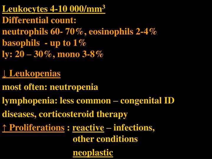
Leukocytes 4-10 000/mm 3 Differential count: neutrophils 60- 70%, eosinophils 2-4% basophils - up to 1% ly: 20 – 30%, mono 3-8%. ↓ Leukopenias most often: neutropenia lymphopenia: less common – congenital ID diseases, corticosteroid therapy

E N D
Leukocytes 4-10 000/mm3Differential count:neutrophils 60- 70%, eosinophils 2-4%basophils - up to 1%ly: 20 – 30%, mono 3-8% ↓ Leukopenias most often: neutropenia lymphopenia: less common – congenital ID diseases, corticosteroid therapy ↑ Proliferations: reactive – infections, other conditions neoplastic
NEOPLASTIC PROLIFERATIONS OF WHITE CELLS • myeloid from the hematopoietic stem cells giving rise to cells of myeloid lineage thrombocytic, granulocytic, erythroid • Acute myelogenous (myeloid) leukaemias • Myelodysplastic syndromes • Chronic myeloproliferative neoplasms B. lymphoid- tumors of lymphocytes, lymphomas and leukaemia • Hodgkin lymphoma versus non-Hodgkin lymphomas; • B- versus T- cell lymphomas • precursor cells (B or T or NK) – lymphoblastic versus mature cells – B or T (or NK) C. histiocytoses – proliferative lesions of histiocytes Langerhans histiocytosis
Myelodysplastic syndrome Bone marrow(BM) hypercellular Dysplastic Ineffective – bone marrow failure Peripheral blood (PB) Blood cells: few and pathological (size, shape, function)
Myelodysplastic syndrome • clonal disorders of stem cells • defects of maturation in the BM - ineffective hematopoiesis (progressive failure of BM function) • cells in the PB : decreased numbers – pancytopenia + defective in function, pathological shapes • BM: hypercellular, • but dysplastic: pathological forms, architecture • blasts may be increased • (but less than 20%, threshold AML versus MDS)
Myelodysplastic syndrome Clinical symptoms and complications ???
Myelodysplastic syndrome Clinical symptoms and complications • Anemia • Thrombocytopenia - bleeding • Leukopenia – infection • (Splenomegaly +-)
Myelodysplastic syndrome purpura
Myelodysplastic syndrome • primary – de novo - old people – over 60 • secondary – therapy related – toxic exposure - worse prognosis
Myelodysplastic syndrome Subcategories • Refractory anemia –– unilineage dysplasia • RA with ringed sideroblasts – the nucleus encircled by siderotic granules • RA with multilineage dysplasia • RA with excess blasts –more than 5%, less than 20% • MDS unclassifiable • MDS assoc. with isolated del. (5q) chromosome
Sideroblastic anaemia (Perl´s stain) collars of iron granules around the nucleus
Chronic myeloproliferative diseases Clonal disorders Adults 1. Chronic myeloid leukemia 2. Polycythemia vera 3. Essential thrombocytemia 4. Chronic osteomyelofibrosis
Chronic myeloproliferative diseases Common principles: 1. Bone marrow: stem cell genetic abnormalities, neoplastic proliferation of one or more (all) BM myeloid series (red, white, megakaryocytes) disorder of an individual series more pronounced in each of the categories 2. Peripheral blood: increased numbers of cells; relatively normal maturation 3. Splenomegaly, hepatomegaly sequestration of excess blood cells, extramedullary hematopoiesis, leukaemic infiltration
Chronic myeloproliferative diseases phases of the disease in time: 1. onset insidious proliferative phase, 2. progression - spent phase - osteomyelofibrosis - blast phase all can (do not have to) progress to AL; CML does it invariably)
Chronic myelogenous leukaemia (CML) t(9; 22) – Philadelphia chromosome, bcr-abl gene pluripotent stem cell defect abnormal fusion protein - increased tyrosine kinase activity most striking : proliferation of G increased cellularity maturation retained (no hiatus leukaemicus) hematopoiesis also extramedullary splenomegaly (hepatomegaly) PB: leukocytosis – exceeds even 100 000/ mm3
CML PB: no hiatus leukaemicus; mature neutrophils, some metamyelocytes, and a myelocyte.
Chronic myelogenous leukaemia (CML) Phases 1. chronic – aver. 3 ys 2.accelerated – gradual failure of response to treatment, increasing anemia and thrombocytopenia, basophilia 3. blast crisis –after accelerated phase or without the acceler. phase Blast crisis = acute leukaemia - 70% myeloid, 30% lymphoblastic
Chronic myeloid leukaemia uric acid deposition
Polycythaemia vera • increased proliferation of all three series • most striking: red cells • hypercellular BM • PB: HTC 60%, Hb over 180g/l
Polycythaemia vera • increased RC mass - symptoms: hypervolemia, blood stasis (mostly venous), cyanosis – stagnation and deoxygenation of blood hypertension, thromboses, bleeding – abnormal blood flow, abnormalities of PLT • granulopoiesis may be elevated • plt elevated + functional abnormalities • SPENT PHASE: myelofibrosis (20%/10 ys)
Polycythemia vera distension of retinal vessels
Polycythemia vera, spent phase, advanced marrow myelofibrosis. Massive splenomegaly (3020 gm; normal: 150 to 200 gm) largely owing to extramedullary hematopoiesis
Essential thrombocythaemia • the least common CMPD • PLT exceed 600 000 /mm3 • BM: increased cellularity, megakaryocytes: abnormal, often large • PB: PLT: often large • Symptoms: • thrombosis and hemorrhage – abnormalities of quantity and quality of PLT • rel. indolent Giant platelets
Essential thrombocythaemia haemorrhages
Essential thrombocythaemia thrombosis, gangrene
Chronic idiopathic myelofibrosis • abnormal neoplastic megakaryocytes release fibrogenic factors – PDGF and TGFa stimulate fibroblasts to proliferation • early: BM hypercellular, minimal fibrosis • progression: BM hypocellular, fibrotic; osteosclerosis • obliteration of BM space: extramedullary hematopoiesis - spleen; later: liver • PB: leukoerythroblastic=erytroid and granulocytic precursors • 20% - progression to AML
Primary myelofibrosis (peripheral blood smear). Two nucleated erythroid precursors and several teardrop-shaped red cells (dacryocytes). Immature myeloid cells present in other fields. An identical picture - in other diseases producing marrow distortion and fibrosis.
Leukaemia • Acute – myeloid; lymphoblastic - B, T • Chronic – myeloid, lymphocytic – B, T
Acute leukaemia (AL) Common: acute course untreated: death in weeks, months Problems – symptoms result from: A. failure of normal hematopoiesis – anemia, neutropenia, thrombocytopenia B. infiltration of organs by neoplastic cells • 1. Myeloid (adults) • 2. Lymphoblastic (young; B or T) further subdivision: genetics, morphology, immunophenotype
Acute myeloid leukaemia • Categories: • AML with recurrent gen. abnormalities –balanced translocations, often complete remission, favourable prognosis (fusion gene - chimeric protein); • t(15;17) – AML M3 – promyelocytic – treatment with transretinoic acid; • t(8; 21) or inversion of chromosome 16 • AML therapy related • AML with multilineage dysplasia • AML – NOS- minim.differentiated • Without maturation • With maturation
Acute myeloid leukaemia • FAB classification = French – American – British • M0 – without maturation – myeloblastic • M1 – without maturation • M2 – with maturation • M3 – promyelocytic – now categorised rather according to the genetics – t(15; 17) • M4 – myelomonocytic • M5 – a – monoblastic, b- monocytic • M6 – erythroid • M7 – megakaryoblastic
Acute myeloblastic leukaemia: Staphylococcus aureus lesions
Acute myeloblastic leukaemia mixed infection Streptococci, Candida previous chemotherapy and neutropenia
AML, M5 leukaemic infiltration of the gums expansion and thickening partial coverings of the teeth
Myeloid sarcoma Tumour mass of immature myeloid cells Extramedullary (bone) Association - before or concurrently: 1. AML (or as a relapse) 2. Chronic myelogenous leukaemia other myeloproliferative disorders 3. MDS Extramedullary myeloid tumour, granulocytic sarcoma, chloroma
Myeloid sarcoma Localization 1. Bones – subperiosteal (skull, paranasal sinuses, sternum, ribs, vertebrae, pelvis) 2. Lymph nodes • Skin
Myeloid sarcoma - poorly differentiated …a high index of suspicion... Stains: CHAE, MPO, lysozyme; CD15, CD68, CD117, CD43 (CD43 only!) Differential diagnosis 1. Lymphoblastoma 2. Burkitt lymphoma 3. Large cell lymphoma 4. Small round cell tumours (neurobl., Ewing/PNET, medullobl.)
Myeloid sarcoma Prognosis If + MPD, MDS = as a blast transformation If + AML – as this AML If isolated: curative radiotherapy prolonged survival
Precursor (lymphoblastic)B-cell neoplasmsB-acute lymphoblastic leukaemia/lymphoblastic lymphoma • 1. Leukaemia (more common): involves the bone marrow and PB • 2. occasionally solid primary nodal or extranodal mass /B-lymphopblastic lymphoma/ without PB and BM involevement - leukaemization possible. biologic unity of B-ALL and B-LBL, division arbitrary
Precursor (lymphoblastic)B-cell neoplasms • Small to medium sized cells • scant cytoplasm, dispersed chromatin, and inconspicuous nucleoli
Acute lymphoblastic leukemia/lymphoma. Lymphoblasts: condensed nuclear chromatin, small nucleoli, and scant agranular cytoplasm
Lymphoblastic leukaemia • More common B • B-ALL – children, but also adults; relatively frequent, good prognosis • Children: 95% complete remission, 80% cured /adults worse/ Lymphoblastoma (LBL) • More common: T • B-LBL – rare T-LBL- rapidly growing mass in mediastinum, adolescent male
Acute B-cell lymphoblastic leukaemia • B-ALL – children, but also adults; relatively frequent • B-LBL - much less common - skin, bone, soft tissues, LN • Symptoms: BM failure • Enlarged LN, liver, spleen • Antigenic profile:TdT, CD10 (CALLA) various degree of differentiation, B-antigens (CD79a, CD20) • Genetic abnormalities– prognostically important • Good: hyperdiploidy; t (12, 21) • Poor: t(9, 22), hypodiploidy • In general: a good prognosis leukaemia • Children: 95% complete remission, 80% cured /adults worse/
Precursor T cell neoplasms • T-ALL– less common than B-ALL • T-ALL, T-LBL - adolescent male • clinically: similarities with B-ALL; • all acute leukemias share some clinical similarities • But difference: AML adults, ALL: children • T-LBL - rapidly growing mass in mediastinum