Download
1 / 31
350 likes | 667 Vues
流式細胞分析儀技術 Flow cytometer analysis and applications on apoptosis. 分細所 張新侯. What is flow cytometer. BD-Calibur . BD FACSVantage. Flow cytometer microscope. Flowing cells Static cells In great number Small amount Subpopulation analysis Cellular signaling. Comparisons

E N D
流式細胞分析儀技術 Flow cytometer analysis and applications on apoptosis 分細所 張新侯
What is flow cytometer BD-Calibur BD FACSVantage
Flow cytometermicroscope Flowing cellsStatic cells In great numberSmall amount Subpopulation analysis Cellular signaling • Comparisons Flow cytometer is a special microscope
Specific functions (1)Multiple characterizations (2)High purity: purity:>99%; (3)High throughput: 150,000cells/ sec
Basic Principles 流體動力學聚焦 Laser beam
laser mirrors 1 2 3 Side scatter or fluorescence Cell Forward scatter Filters PMT lance Light path
Data analysis Light signals FSC SSC FL1 FL2 FL3 Electric signals log linearlinearlinearlog Data recording
Non-fluorescent signal 颗粒度 细胞大小
Fluorescent signals 螢光染料被激發而發射的光信號。 定量染色 螢光信號大小 被標記组分含量的定量 多螢光標記胞内多種组分,實現多參數测量
PI+Annexin-V细胞凋亡檢測 DNA含量-PI Annexin-V
laser Cell sorting
流式細胞儀的測量對象 • 大小 懸浮在溶液中的相互離散顆粒。 大小範圍:0.2uM-300uM。 • 對象類型 细胞類型: (1)高等真核细胞; (2)酵母; (3)细菌; (4)多細胞的聚集體,如胰島素表現細胞等。 非生命顆粒: 細胞核、染色體、和其它胞器以及顆粒等。
流式细胞儀能够檢测的细胞屬性和成份舉例 1)不需染色: 大小,細胞質顆粒度,细胞自發螢光,色素,细胞精確計數。 2)需染色或標記: 细胞成分: 细胞膜:流動性,通透性,膜電位 细胞内離子:H+(可檢測PH值),Na+,K+,Ca2+(结合的 和自由的)。 核酸:DNA含量,DNA成分,DNA合成,RNA,染色 體結構等 總蛋白質 蛋白質分子相相互作用 抗原 细胞骨架成分 受體
功能屬性: 酶活性 细胞凋亡 细胞壞死 细胞活性 氫硫基 凝血素结合位置 藥物動力学 …………………….. 經驗談: 凡细胞内能進行螢光標記的成分或變化,都可用流式细胞儀進行檢測。
细胞周期分析 细胞數 DNA 含量 G1 S phase M G0/1 G2 G1 G2/M S 1 2 1 DNA含量 细胞周期
染色體测定 2 1 4 3 白血病患者 5 1 / 3 6 Hoechst 33258 (AT) X 8 7 recip 9-22 13 Y 18 9-12 14 15 16 17 20 21 19 22 Ph Chromomycin A3 (GC)
應用: 範例 登革 Project -淋巴球分析
How to define cell cycle stage? Propidium iodide (PI) staining and flow cytometer analysis. Quiescence Sub-G1 phase
DNA ladder DNA fragmentation staining with Hoechst
Terminal deoxynucleotide transferase dUTP Nick End Labeling (TUNEL) When DNA strands are cleaved or nicked by nucleases, 3’-hydroxyl ends are exposed. The hydroxyl groups can then serve as a starting point for terminal deoxynucleotidyl transferase (TdT), which adds deoxyribonucleotide in a template-independent fasion. Addition of BrdUTP to the TdT reaction thus provides a means to label the DNA strand breaks. Once incorporated into DNA, the BrdU can be detected by an anti-BrdU antibody using standard immunohistochemical techniques.
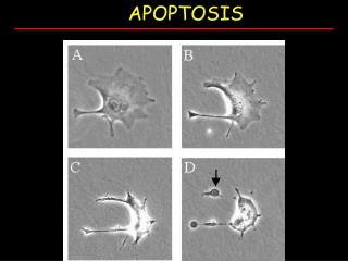
Phosphatidylserine ExposureAnnexin V Staining In viable cells, phosphatidylserine (PS) is located on the cytoplasmic surface of the cell membrane. As cells undergo apoptosis, PS is translocated to the outer leaflet of the plasma membrane and exposed to the extracellular environment. The human vascular anticoagulant, annexin V, is a 35-36 kD Ca2+-dependent phospholipid-binding protein that has a high affinity for PS. Thus, annexin V labeled with a fluorophore can identify apoptotic cells by binding to PS exposed to the outer leaflet.
Analysis under fluorescence microscope Analysis under flow cytometer