Download
1 / 18
340 likes | 1.19k Vues
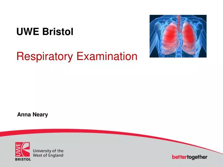
UWE Bristol Respiratory Examination. Anna Neary. Respiratory Examination. Introduction

E N D
UWE BristolRespiratory Examination Anna Neary
Respiratory Examination Introduction This activity looks at a systematic approach to respiratory examination. You will need to have completed learning activities consultation models and history taking before undertaking this learning activity. In this activity, you will: • Discover the anatomy of the thorax and lungs • Consider a systematic approach to respiratory examination including inspection, palpation and auscultation • Consider the elements of a general survey • Identify some normal and abnormal breath sounds
Respiratory Examination Anatomy The respiratory system consists of the airways, lungs, bony thorax and respiratory muscles all working together . The Lungs are covered in a lining called the visceral pleura. There are three lobes on the right side of the chest (upper, middle and lower). There are two lobes on the left side of the chest (upper and lower) to accommodate the heart. The lower airways start with the trachea and then sub divide into the right and left main bronchi. These bronchi divide into lobar bronchi, then secondary bronchi, tertiary bronchi, terminal bronchioles , respiratory bronchioles and finally alveolar ducts. The bronchioles sub divide into several alveolar ducts, which in turn will have a number of alveolar sacs. The alveoli consists of alveolar cells, which are simple squamous epithelial cells, this is the main site for gas exchange. Anatomy of the chest wall, the chest consists of 12 ribs with intercostal spaces, a sternal angle adjacent to 2nd rib, costal cartilage from rib 1-7 articulate with sternum. Ribs 8-10 articulate with the costal cartilage just above them and ribs 11 and 12 have no anterior attachments.
Respiratory Examination Anterior axillary line Anterior axillary line Vertebral line Mid clavicular line Sternal line Mid-axillary line Left scapular line Posterior axillary line Use the vertical anatomical lines as shown above to describe your findings.
Respiratory Examination Inspiration and expiration Normal inspiration uses the diaphragm that goes down and flattens, this is an active process. The intercostal muscles raise the sternum and ribs and in turn air enters the lungs. Negative alveolar pressure is created. Air moves into the lungs until the pressures are equal. Expiration is a passive process where the intercostal muscles and diaphragm relax. The lungs recoil to their resting size and position. Positive alveolar pressure is created and the air moves out of the lungs.
Respiratory Examination Systematic Examination There are three aspects to consider when carrying out a systematic examination: LOOK: inspect FEEL: palpate and percuss LISTEN: auscultate
Respiratory Examination General Survey First you need to take time to look at your patients overall appearance, remembering the importance of basic assessment i.e. airway, breathing, circulation, disability and exposure and move on only when there is no immediate intervention in these areas. Also consider whether your patient appears to be alert or confused or aggressive. You will need to check their blood pressure in both arms, heart rate, respiratory rate and pulse. Look at hands, check for signs of cyanosis and any nail deformities. Does you patient look pale, sweaty are there any signs of cyanosis, are there signs of any shortness of breath?Examining the hands is very important in patients with respiratory disease as there are many abnormalities that can be found such as finger clubbing, peripheral cyanosis, tobacco staining and CO2 retention. Other signs and symptoms of a respiratory condition include, shortness of breath, anxious or distressed, pallor, cough, wheezing, confusion, stridor, cyanosis of lips and peripheral cyanosis, chest pain, tripod sitting position, clubbing of finger nails, fine hand tremor when asked to place hands out in front of them for 20-30 seconds.
Respiratory Examination Inspection • Check the patients rate, rhythm and depth/work of breathing. • Check position of trachea, is it midline? • Look at the patients chest (anterior and posterior), does the shape appear normal? • Is there swelling, bruising or scarring on the chest wall? • Can you hear any abnormal noises (without stethoscope) i.e. grunting, wheeze, stridor? • Inspect Sputum.
Respiratory Examination Sputum There are four main types of sputum; Serous- This is clear and watery although can be frothy and pink in colour. It can indicate acute pulmonary oedema and can be a sign of alveolar cell cancer. Mucoid- This is clear grey or white in colour and is viscid in texture. It can indicate the presence of chronic bronchitis, COPD or asthma. Purulent- This is usually yellow or green in colour. The yellow purulent sputum indicates acute bronchopulmonary infection and /or asthma. Green purulent sputum indicates a long standing infection such as pneumonia, bronchiectasis or cystic fibrosis or the presence of a lung abscess Rusty- This is as the name suggests rusty red in colour and indicates the presence of pneumoccal pneumonia.
Respiratory Examination Palpate the anterior and posterior chest wall, check for tenderness.
Respiratory Examination Chest Expansion it is important when examining the chest to check for symmetry when the patient is breathing. To do this place your hands at the level of the 10th rib as shown in the photograph. Ask the patient to breath in deeply, watch your thumbs move apart, this should be equal. Also feel the rib cage expand and contract. This can be done on the anterior of the chest or the posterior
Respiratory Examination Tactile Fremitus Fremitus refers to the palpable vibrations transmitted through the bronchopulmonary tree to the chest wall when the patient speaks. To detect tactile fremitus you need to place either the ball or bony aspect of your hand or the ulna border of your hand in certain places on the chest and ask the patient to say “99”. You will feel the vibrations through the lungs as air is transmitted. You should perform this anteriorly and posteriorly. • Intense vibrations can indicate consolidation in tissue. • Faint or absent vibrations can indicate obstruction or fluid filled pleural space.
Respiratory Examination Hand positioning for tactile fremitus
Respiratory Examination Percussion Percussion is performed to establish whether the lungs are filled with air, fluid or solid material and to establish lung boundaries. Percuss in the rib spaces, place your middle finger flat in the space and using your dominant hand tap the middle finger with the tip of your finger. Use a systematic approach comparing sides as shown in the picture. Normal percussion sound is resonant, long and loud, low pitched and hollow. A solid area will sound dull and “thud-like”. Hyperinflated lung as in pneumothorax will sound hyperresonant, very loud and lower pitched.
Respiratory Examination Auscultation The sound heard through the stethoscope when you are listening to the chest are different depending on where you are listening. Vesicular sounds are heard over most of the lung fields, this is soft and of a low pitch. Bronchovesicular sounds are heard over the lower trachea and left and right main bronchi, this sound is intermediate in intensity and pitch. Bronchial sounds are heard over the trachea in the neck and they are loud and high pitch. You should listen to the chest in the same pattern as when you percussed the chest, ensuring you compare sides.Thereare many breath sounds that can indicate abnormalities within the lungs. Click on the link and then listen to both recordings of lung sounds.
Respiratory Examination Summary You have now reached the end of this activity. Here is a summary of the key points: • It is important to know the anatomy of the thorax and lungs prior to any examination of the respiratory system. • When you examine the chest use a systematic approach i.e. general survey, inspection, palpation and auscultation. • It is important that you have an understanding of the normal sounds when percussing the chest and the normal breath sounds when ausultating, and normal vibrations when performing tactile fremitus.
Respiratory Examination References • Douglas, G, Nicol, F and Robertson, C (2009) Macleod’s Clinical Examination. Churchill Livingstone • Hogan-Quigley, B, Louise Palm, M, Bickley, L (2012)Bates’ Nursing Guide to Physical Examination and History Taking. First Edition. Lippincott, Williams and Wilkins. • Rushforth, H (2009) Assessment made incredibly easy.First UK Edition. Lippincott, Williams and Wilkins • Tortora, G and Derrickson, B (2010) Essentials of anatomy and physiology. Wiley Plus
Respiratory Examination Guided learning (Duration: 2 hours) • Identify a patient in practice and under the supervision of an Advanced Nurse Practitioner or doctor, watch a respiratory examination and then perform a respiratory examination. Use the templates provided as an aid memoir. • Discuss your findings with the ANP or doctor. • Research common respiratory conditions such as: • COPD • Asthma The British Thoracic website will help your research. • Write a reflective piece of work about a patient you have seen and the guidelines you have learnt. • You will also be able to attend a day long face to face skills workshop to complement this learning activity.