Download
1 / 40
420 likes | 662 Vues
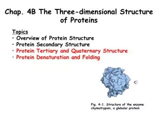
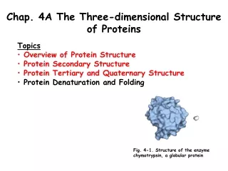
Chapter Four The Three-Dimensional Structure of Proteins. Protein Structure. Many _________________ are possible for proteins: Due to flexibility of amino acids linked by peptide bonds

E N D
Protein Structure • Many _________________ are possible for proteins: • Due to flexibility of amino acids linked by peptide bonds • At least one major _________________ has biological activity, and hence is considered the protein’s _______________ _________________
Levels of Protein Structure ___ structure: the ________________ of amino acids in a polypeptide chain, read from the N-terminal end to the C-terminal end ___ structure: the ______________ ______________ arrangements (conformations) in localized regions of a polypeptide chain; refers only to interactions of the peptide backbone e. g., -helix and -pleated sheet ___ structure: 3-D arrangement of all atoms ___ structure: arrangement of monomer subunits with respect to each other
1˚ Structure • The 1˚ sequence of proteins determines its 3-D conformation • Changes in just one amino acid in sequence can alter biological function, e.g. hemoglobin associated with sickle-cell anemia • Determination of 1˚ sequence is routine biochemistry lab work (See Ch. 5).
2˚ Structure • 2˚ of proteins is hydrogen-bonded arrangement of _________________ of the protein • Two bonds have _____________ ____________: • Bond between _________________ and _________________ in residue • Bond between the _________________ and _________________ of residue • See Figure 4.1
-Helix • Coil of the helix is ___________ or _____________ • There are ______ amino acids per turn • Repeat distance is _____ Å • Each peptide bond is _________ and ________ • C=O of each peptide bond is _________________ _________ to the N-H of the fourth amino acid away • C=O----H-N hydrogen bonds are ________________ helical axis • All R groups point _________________ from helix
-Helix (Cont’d) • Several factors can _______________ an -helix • _______ creates a bend because of (1) the restricted rotation due to its cyclic structure and (2) its -amino group has no N-H for hydrogen bonding • strong _________________ _________________ caused by the proximity of several side chains of like charge, e.g., Lys and Arg or Glu and Asp • _________________ _________________ caused by the proximity of bulky side chains, e.g., Val, Ile, Thr
-Pleated Sheet • Polypeptide chains lie adjacent to one another; may be _________________ or _________________ • R groups ________, first above, then below _______ • Each peptide bond is ________ and ________ • C=O and N-H groups of each peptide bond are ____________ to axis of the sheet • C=O---H-N hydrogen bonds are between adjacent sheets and ____________ to the direction of the sheet
-Pleated Sheet (Cont’d) -bulge- a common nonrepetive irregular 2˚ motif in ____________ structure
Structures of Reverse Turns • ____________ found in reverse turns • Spatial (steric) reasons • Polypeptide changes direction • ____________ also encountered in reverse turns. Why?
-Helices and -Sheets • __________________ structures: the combination of - and -sections, as for example • b unit: two parallel strands of -sheet connected by a stretch of -helix • unit: two antiparallel-helices • -meander: an antiparallel sheet formed by a series of tight reverse turns connecting stretches of a polypeptide chain • Greek key: a repetitive supersecondary structure formed when an antiparallel sheet doubles back on itself • -barrel: created when -sheets are extensive enough to fold back on themselves
Collagen Triple Helix • Consists of three polypeptide chains wrapped around each other in a ropelike twist to form a triple helix called __________________; MW approx. 300,000 • 30% of amino acids in each chain are Pro and Hyp (hydroxyproline); hydroxylysine also occurs • Every third position is Gly and repeating sequences are X-Pro-Gly and X-Hyp-Gly • Each polypeptide chain is a helix but not an a-helix • The three strands are held together by hydrogen bonding involving hydroxyproline and hydroxylysine • With age, collagen helices become cross linked by covalent bonds formed between Lys and His residues
Fibrous Proteins • Fibrous proteins: contain polypeptide chains organized approximately parallel along a single axis. They • consist of long fibers or large sheets • tend to be mechanically strong • are insoluble in water and dilute salt solutions • play important structural roles in nature • Examples are • ____________ of hair and wool • ____________ of connective tissue of animals including cartilage, bones, teeth, skin, and blood vessels
Globular Proteins • Globular proteins: proteins which are folded to a more or less spherical shape • they tend to be soluble in ____________ and ____________ solutions • most of their polar side chains are on the outside and interact with the aqueous environment by hydrogen bonding and ion-dipole interactions • most of their nonpolar side chains are ______ ______ • nearly all have substantial sections of _____________ and ____________
3˚ Structure • The ____________ arrangement of atoms in the molecule. • In ____________ protein, backbone of protein does not fall back on itself, it is important aspect of 3˚ not specified by 2˚ structure. • In ____________ protein, more information needed. 3k structure allows for the determination of the way helical and pleated-sheet sections fold back on each other. • Interactions between ______ ______ also plays a role.
Forces in 3˚ Structure • _________ interactions, including • _________ _________ between polar side chains, e.g., Ser and Thr • _________ interaction between nonpolar side chains, e.g., Val and Ile • _________ _________ between side chains of opposite charge, e.g., Lys and Glu • _________ _________ between side chains of like charge, e.g., Lys and Arg, Glu and Asp • _________ interactions: Disulfide (-S-S-) bonds between side chains of _________
3° and 4° Structure • Tertiary (3°) structure: the arrangement in space of all atoms in a polypeptide chain • it is not always possible to draw a clear distinction between _________ and _________ structure • Quaternary (4°) structure: the association of polypeptide chains into _________ • Proteins are divided into two large classes based on their three-dimensional structure • _________ proteins • _________ proteins
Determination of 3° Structure • X-ray crystallography • uses a perfect crystal; that is, one in which all individual protein molecules have the same 3D structure and orientation • exposure to a beam of x-rays gives a series of diffraction patterns • information on molecular coordinates is extracted by a mathematical analysis called a Fourier series • 2-D Nuclear magnetic resonance • can be done on protein samples in aqueous solution
X-Ray and NMR Data High resolution method to determine 3˚ structure of proteins (from crystal) Diffraction pattern produced by electrons scattering X-rays Series of patterns taken at different angles gives structural information Determines solution structure Structural info. Gained from determining distances between nuclei that aid in structure determination
Myoglobin • A single polypeptide chain of ____ amino acids • A single ______ group in a _____________ pocket • 8 regions of -helix; no regions of -sheet • Most _______ side chains are on the __________ • ________ side chains are folded to the __________ • Two His side chains are in the interior, involved with interaction with the heme group • Fe(II) of heme has 6 coordinates sites; 4 interact with N atoms of heme, 1 with N of a His side chain, and 1 with either an O2 molecule or an N of the second His side chain
Denaturation • Denaturation: the loss of the structural order (2°, 3°, 4°, or a combination of these) that gives a protein its biological activity; that is, the loss of biological activity • Denaturation can be brought about by • heat • large changes in pH, which alter charges on side chains, e.g., -COO- to -COOH or -NH+ to -NH • detergents such as sodium dodecyl sulfate (SDS) which disrupt hydrophobic interactions • urea or guanidine, which disrupt hydrogen bonding • mercaptoethanol, which reduces disulfide bonds
Denaturation and Refolding in Ribonuclease Several ways to denature proteins • Heat • pH •Detergents • Urea • Guanadine hydrochloride
Quaternary Structure • Quaternary (4°) structure: the association of polypepetide ________ into _____________ proteins • dimers • trimers • tetramers • Noncovalent interactions • electrostatics, hydrogen bonds, hydrophobic
Oxygen Binding of Hemoglobin (Hb) • A _________ of two -chains (141 amino acids each) and two -chains (153 amino acids each); a2b2 • Each chain has 1 heme group; hemoglobin can bind up to 4 molecules of O2 • Binding of O2 exhibited by _________ ___________; when one O2 is bound, it becomes easier for the next O2 to bind • The function of hemoglobin is to transport oxygen • The structure of oxygenated Hb is different from that of unoxygenated Hb • H+, CO2, Cl-, and 2,3-_______________ (BPG) affect the ability of Hb to _________ & ________ oxygen
Conformation Changes That Accompany Hb Function • Structural changes occur during binding of small molecules • Characteristic of __________________ behavior • Hb exhibits different 4˚ structure in the bound and unbound oxygenated forms • Other _________ are involved in cooperative effect of Hb can affect protein’s affinity for O2 by altering structure
Protein Folding Dynamics •Can 3˚ structure of protein be predicted? Yes, within limitations • The integration of biochemistry and computing has led to bioinformatics • Protein structure prediction is one of the principal application of bioinformatics • First step to predict protein structure is to search for sequence homology
Hydrophobic Interactions • Hydrophobic interactions are major factors in protein folding • Folds so that nonpolar hydrophobic side chains tend to be on inside away from water, and polar side chains on outside accessible to aqueous environment • Hydrophobic interactions are __________________
Protein Folding Chaperones • In the protein-dense environment of a cell, proteins may begin to fold _________ or may associate with other proteins before folding is completed • Special proteins called _________ aid in the correct and timely _________ of many proteins • hsp70 were the first chaperone proteins discovered • Chaperones exist in organisms from prokaryotes to humans