Download
1 / 34
540 likes | 1.36k Vues
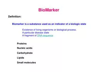
BioMarker. Definition:. Biomarker is a substance used as an indicator of a biologic state Existence of living organisms or biological process. A particular disease state A fragment of DNA sequence. Proteins. Nucleic acids. Carbohydrate. Lipids. Small molecules. Biomarkers.

E N D
BioMarker Definition: Biomarker is a substance used as an indicator of a biologic state Existence of living organisms or biological process. A particular disease state A fragment of DNA sequence Proteins Nucleic acids Carbohydrate Lipids Small molecules
Biomarkers Location of the biomarker: Location of a particular molecule can also be a marker Cellular-subcellular locations Tissue or organ locations Thymidylate synthase (TS) nucleus vs the cytoplasm The higher the level in NE, lower level in the cyto. Lower survival in colorectal cancer patients Galectin-3 Galectin-3 is a member of lectin family. It is wildly distributed In tissue of epithelial, fibroblast, and dendritic cells. Blood Galectin-3 Predict the outcomes for patients with symptoms of heart failure
Biomarker Detection of biomarker Detection of biomarker – diagnosis Self properties, e.g enzymatic activities Antibodies, IHC, ELISA Detection of biomarker Quantitative a link between quantity of the marker and disease Qualitative a link between exist of a marker and disease
Ideal Marker for diagnosis Biomarker & Diagnosis Should have great sensitivity, specificity, and accuracy in reflecting total disease burden. A tumor marker should also be prognostic of outcome and predictive of tumor recurrence and effectiveness of anti-cancer treatments. Biomarker for Screening • The marker must be highly specific, minimize false positive and negative • The marker must be able to clearly reflect the early stage of disease • The marker must be easily detected without complicated medical • procedures. The disease markers released to serum and urine are good • targets for application of early screening. • The method for screening should be cost effective. Samples for biomarker detection Blood, urine, or other body fluids samples Tissue samples
Biomarker Discovery and Validation Correlation: a biomarker vs a disease or status of a disease Do not need understand functions Detection: Detection of a particular marker is important Validation: Build statistical correlation – large number of samples Validation: sensitivity and specificity Validation: Stand alone vs along with other markers
Common Biomarker for Cancer AFP (>100 ng/ml) Alpha fetoprotein (AFP) is typically found in the developing fetus. Because of the association of the rapid cell growth, this fetal protein is also used as a tumor marker. non cancerous liver diseases such as cirrhosis and viral hepatitis can lead to high level AFP CEA (>10 ng/ml) Carcinoembryonic antigen (CEA) is produced during fetal development. After birth, the production of CEA stops and is undetectable. CEA has also been found elevated in nonmalignant tumors such as pleural effusions. Elevation of CEA after conventional treatment of neoplasms has been correlated with a recurrence of cancer CA19-9 (>37 U/ml) Carbohydrate Antigen 19-9 (CA 19-9) is present in the fetus in the epithelium of the fetal stomach. It is primarily used as a marker for pancreatic cancer. High levels also exist in conditions such as non-malignant liver disease and other disorders of the gastrointestinal tract.
Common Biomarker for Cancer hTG (>10 ng/ml) Thyroglobulin (hTG) is a glycoprotein that is found in thyroid gland. This protein binds to thyroxine, which controls the rate of metabolic processes in body. HCG (>10 mIU/ml) Human Chorionic Gonadotropin-beta (HCG) is normally produced by the placenta during pregnancy, an indicator of pregnancy. The protein can be detected in serum or urine. Non-malignant elevations may be observed in pregnancy, ulcers, duke’s disease, and cirrhosis. Levels of HCG are useful in monitoring the effectiveness of treatment. Ferr (>120 ng/ml) Ferritin is an iron binding storage found in the liver, spleen, and bone marrow. Elevated levels observed in non-cancerous conditions include rheumatoid arthritis and anemia
Common Biomarker for Cancer NSE (>12 ng/ml) Neuron Specific Enolase (NSE) is produced by neurons and neuroendocrine cells of the central and peripheral nervous system. b2M (high level) Beta 2-Microglobulin, (B2M), is an 11 kD protein associated with the outer membrane of many cells including lymphocytes. It is the small subunit of the MHC class I molecule. B2M is present in small amounts in serum, csf, and urine of normal people. A non-malignant condition associated with high b2M levels is pancreatitis a2M (<500 mg/ml) Alpha 2 Macroglobulin (A2M) is a large protease inhibitor, 720 kD, capable of irreversibly binding and inhibiting a wide variety of proteases including plasmin, pepsin, trypsin, chymotrypsin, and cathepsin D.
Prostate Cancer marker PSA PSA is a protein normally made in the prostate gland in ductal cells that make some of the semen. PSA helps to keep the semen liquid. PSA, also known as kallikrein III, seminin, semenogelase, γ-seminoprotein and P-30 antigen, is a glycoprotein, a serine protease PSA has several forms in serum Free Pre-enzyme Complex Inactive
Prostate Cancer Diagnosis with PSA Cancer of the prostate does not cause any symptoms until it is locally advanced or metastatic. There is a correlation between elevated PSA and prostate cancer. Diagnosis of PSA for prostate cancer in the most time means measure the PSA In serum samples. Detection of PSA allow early detection of prostate cancer. Large screening trials have shown that PSA nearly doubles the rate of detection possible by combine with other methods. Based on these data, PSA testing was approved by the US FDA for the screening and early detection of prostate cancer. PSA is also found in the cytoplasm of benign prostate cells. Separately measure different forms of PSA give better results
Prostate-specificmembraneantigen (PSMA) is a type 2 integral membrane glycoprotein. PSMA possesses glutamate carboxypeptidase activity, Detectable levels of PSMA are also found in small intestine and brain. Intestinal PSMA may play a role in the metabolism of dietary-glutamated folate derivatives. Brain PSMA, also referred to N-acetylated -linked acidic dipeptidase (NAALADase), may modulate glutamatergic neurotransmission Prostate Marker PSMA In addition to being abundantly and preferentially expressed on the surface of prostate cancer cells, PSMA is also present on endothelial cells of new blood vessels PSMA also expressed in brush border of proximal tubules of normal kidneys and tissue samples from both benign and malignant renal lesions (neovasculature) The role of PSMA as a cancer marker is still under investigation. PSMA is sensitive to reflect prostate cancer. The specificity, however, is not fully verified. Most use is image the prostate metastasis by radio-isotopes
Prostate Marker PSMA The role of enzymatic activity (folate hydrolase) of PSMA in cellular transformation and metastasis is not understood. PSMA has isoforms produced by alternative splicing. In normal prostate epithelia, PSMA is expressed primarily as a cytoplasmic protein termed PSM' In prostate carcinomas, however, differential mRNA splicing leads to expression of PSMA as a type 2 integral membrane glycoprotein possessing a 19-aa cytoplasmic fragment, a single 24-aa membrane-spanning domain, and a 707-aa extracellular region Unlike PSA, PSMA is not a secret protein. Serum level is very low even elevated. PSMA is membrane protein, ideal for targeting
Ovarian Cancer Diagnosis by CA-125 CA-125, a product of the MUC16 gene, is a mucin made by certain cells in the body which include those of the uterine tubes, uterus, cervix, and the lining of the abdominal and chest cavities CA125 is a membrane glycoprotein that has very short cytoplasmic domain and a very long extracellular domain. The cellular function of the protein is still unknown. The protein exist in the cells of normal and cancerous tissues of ovarian. The release of proteolytic fragments of CA125 leads to elevation of CA125 level in bloodstream, which is associated with progression of ovarian cancer and a few other cancer types. The CA125 level can be over 10 folds higher in ovarian cancer patients compared to the level in normal. Diagnosis of ovarian cancer is often simple. A pelvic mass is suspicious and is very often associated with ovarian cancer. However, when the pelvic mass is detected, the ovarian cancer usually has already advanced to quit advanced stage. There is no symptom for ovarian cancer, especially during early stage.
Her-2/neu The HER (erbB) family member are Growth factor receptors. In the HER (erbB) family, only Her-2 did not have identified ligand. Expression level correlates with growth of breast cancer cells. Diagnostic/prognostic applications. HER2-positive metastatic breast cancer have a more aggressive disease, greater likelihood of recurrence, poorer prognosis and decreased survival.
Beta-2-microglobulin (b2M) B2M is the small subunit of the MHC class I molecule. How does the protein promotes tumor growth and metastasis – the expression of MHC class 1 in many tumor cells are repressed. An hypothesis is that b2M is a signaling molecule that linked to cell growth and survival pathway. What are the target of the signal? Receptors? Known or unknown pathway?
Biomarker & Targeted Imaging Imaging probe link to a targeting moiety that target to bio-marker Basic requirement: the marker must presented on cell surface in high number Ideally: Theinteraction between marker and targeting moiety is strong The interaction is highly specific The target internalization – high payload Distribution at neovasculature, minimal tissue penetration
Imaging Targeting PSMA Antibody is targeting moiety Antibody conjugated quantum dots Antibody
Imaging Targeting PSMA Antibody conjugated to radio-labels Patient Images No resolution – hardly tissue image High sensitivity – good for detecting metastasis
MR Image Target Her-2 Advantage for both Her-2 and PSMA imaging is that the high number and distribution of the marker in agiogenesis tissue Disadvantage for both Her-2 and PSMA imaging is that the ligand for Her-2 and PSMA are not identified. Antibodies are the only choice The levels of both Her-2 and PSMA correlate well with aggressiveness of disease
Therapies Target Biomarkers Simply target the biomarker by antibody Therapeutic agent link to a targeting moiety that target to bio-marker Basic requirement: the marker must presented on cell surface Ideally: The interaction is highly specific. The target internalization, accumulation good # of agent. Distribution at neovasculature, minimal tissue penetration. The level of marker correlate with disease progression
Therapy Target Her-2 Herceptin Herceptin binds to HER2-positive cancer cells and may block them from dividing and growing. Herceptin attaches to the HER2-positive cancer cells and may signal the body's immune system to destroy the cell. Herceptin can also conjugated with chemotherapy (paclitaxel) to destroy HER2-positive cancer cells.
Herceptin 100% 33% 10% Mouse Fused Humanized
Therapy Target Her-2 Small Molecules target HER-2 erlotinib (Tarceva), a kinase inhibitor for EGFR/HER1 Target HER-2 Affibody target HER-2 Polypeptides of 58 aa derived from IgG binding domain of protein A, forming a three-helix bundle.
Therapy Target PSMA One important advantage is the quick endocytosis of the PSMA upon the antibody binding. Due to main neovascular distribution, targeting of PSMA is very easy, less tissue penetration. Both Her-2 and PSMA have a good property is their high level correlate well with aggressiveness of disease.
PSMA Target for Tumor Blood Vessel PSMA is found highly expressed in neovascular of many cancer types, especially the new vessel in cancer mass. PSMA is an excellent target for delivery to many cancer
Imaging and Therapeutic Agent The most significance of the double agent is the immediately follow the effectiveness of treatment. Also follow whether the therapies have been targeted to the sites
Diagnosis by Proteomic No single marker can accurately reflecting a disease. Not even a panel of markers. We need analyses a large number of disease markers.