Download
1 / 46
460 likes | 772 Vues
The Cardiovascular System: Blood Vessels. Chapter 20. Introduction. The blood vessels of the body form a closed delivery system that begins and ends at the heart

E N D
The Cardiovascular System:Blood Vessels Chapter 20
Introduction • The blood vessels of the body form a closed delivery system that begins and ends at the heart • Often compared to a plumbing system, it is a far more dynamic system of structures that pulse, constrict and relax and even proliferate to meet changing body needs
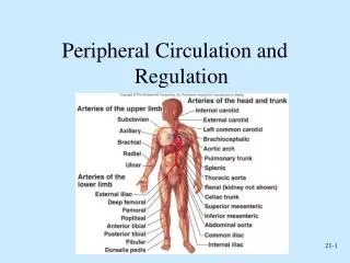
Blood Vessel Structure & Function • The major types of blood vessels are • Arteries • The large distributing vessels that bring blood to the body • Capillaries • The tiny vessels that distribute blood to the cells • Veins • The large collecting vessels that bring blood back to the heart • Intermediate vessels connect • Arterioles bring blood to the capillaries • Venules drain blood from the capillaries
Blood Vessel Structure & Function • The pattern of distribution starts with arteries to arterioles to capillaries to venules to veins • The blood vessels in the adult human body carry blood in a distribution network that is approximately 60,000 miles in length • Only capillaries come into intimate contact with tissue cells and serve cellular needs
Blood Vessel Walls • The walls of blood vessels are composed of three distinct layers or tunics • The tunics surround a central opening called a lumen
Blood Vessel Walls • The innermost tunic is the tunica intima • This tunic contains the endothelium, the simple squamous endothelium that lines all vessels • Its flat cells fit closely together, forming a slick surface that minimizes friction as blood moves through the vessel lumen Tunica adventitia
Blood Vessel Walls • In blood vessels larger than 1 mm in diameter, a sub- endothelial layer of loose connective tissue (a basement membrane) supports the endothelium
Blood Vessel Walls • The middle tunic, the tunica media is mostly circularly arranged smooth muscle cells and sheets of elastin • The activity of the smooth muscle is regulated by vasomotor nerve fibers of the sympathetic division of the autonomic nervous system Tunica media
Blood Vessel Walls • Depending on the needs of the body, the vasomotor fibers can cause vaso-constriction or vasodilation • The activities of the tunica media are critical in regulating circulatory dynamics • Generally, the tunica media is the bulkiest layer in arteries, which bear the chief responsibility for maintaining blood pressure and continuous blood circulation
Blood Vessel Walls • The outermost layer of a blood vessel is the tunica adventitia • This tunic is composed largely of loosely woven collagen fibers that protect blood vessels and anchor it to surrounding structures Tunica adventitia
Blood Vessel Walls • The tunica adventitia is infiltrated with nerve fibers and lymphatic vessels and, in larger vessels, a system of tiny blood vessels • These vessels, the vasa vasorum nourish the external tissues of the blood vessel wall Tunica adventitia
Elastic (Conducting) Arteries • Elastic arteries are thick walled arteries near the heart - the aorta and its major branches • These arteries are the largest in diameter and the most elastic • A large lumen allows them to serve as low resistance pathways that conduct blood from the heart to medium-sized arteries and thus are called conducting arteries
Elastic (Conducting) Arteries • The elastic arteries contain more elastin than any other type of vessel • While present in all three layers, the tunica media contains the most • The abundant elastin enables these arteries to withstand and smooth out large pressure fluctuations by expanding when the heart forces blood into them and then recoiling to propel blood onward into the circulation when the heart relaxes
Elastic (Conducting) Arteries • Elastic arteries also contain substantial amounts of smooth muscle, but they are relatively inactive in vasoconstriction • Because elastic arteries expand and recoil passively to accommodate changes in blood volume, the blood is kept under pressure • Thus, blood flows continuously rather than starting and stopping with each heart beat
Muscular (Distributing) Arteries • The muscular distributing arteries deliver blood to specific body organs and account for most of the named arteries • Proportionately, they have the thickest media of all vessels • Their tunica media contains relatively more smooth muscle and less elastic tissue than that of elastic arteries • They are more active in vasoconstriction and are less distensible
Arterioles • Arterioles have a lumen diameter from 0.3 mm to 10 m, and are the smallest of the arteries • Larger arterioles exhibit all three tunics, but their tunica media is chiefly smooth muscle with a few scattered muscle fibers • The smaller arterioles that lead into capillary beds, are little more than a single layer of smooth muscle cells spiraling around the endothelial lining
Capillaries • The microscopic capillaries are the smallest blood vessels • In some cases, one endothelial cell forms the entire circum- ference of the capillary wall • The average length of a capillary is 1 mm and the average diameter is 8-10 m
Capillaries • Capillaries have a lumen just large enough for blood cells to slip through in single file • Most tissues have a rich supply, but there are a few exceptions • Tendons and ligaments • Cartilage • Epithelia • Cornea
Capillaries • Given their location and the thinness of their walls capillaries are ideally suited for their role of providing access to nearly every cell • Exchange of materials • Gases • Nutrients • Hormones
Types of Capillaries • Structurally there are three types of capillaries • Continuous • Fenestrated • Sinusoidal
Continuous Capillaries • Continuous capillaries abundant in the skin and muscles are the most common • They are continuous in the sense that their endothelial cells provide an uninterrupted lining • Adjacent cells are joined laterally by tight junctions • However, these are usually incomplete and leave gaps of unjoined membrane called intracellular clefts that are just large enough to allow limited passage of fluids
Fenestrated Capillaries • Fenestrated capillaries are similar to the continuous variety except that some of their endothelial cells are riddled with oval pores or fenestrations Intercellular clefts
Fenestrated Capillaries • The fenestrations is usually covered by a thin diaphragm but this variety has much greater permeability to fluids and small solutes • Fenestrated capillaries are found where active capillary absorption or filtrate formation occurs • Fenestrated capillaries of digestive tract • Receive digested food nutrients • Fenestrated capillaries perpetually open • Kidneys for rapid filtration of blood plasma
Sinusoidal Capillaries • Sinusoids are highly modified, leaky capillaries found in only in certain organs (liver, bone marrow, lymphoid tissues, and some endocrine organs) • Sinusoids have large, irregularly shaped lumens and are usually fenestrated • Their endothelial lining is modified to exhibit fewer tight junctions and larger intercellular clefts
Sinusoidal Capillaries • These structural modifications allow large molecules (proteins) and even blood cells to pass between the blood and the surrounding tissues • In the liver, the endothelium of the sinusoids is discontinuous and large macrophages called Kupffer cells form part of the lining • In organs such as the spleen, phagocytes located outside the sinusoid stick cytoplasmic extensions through the inter-cellular clefts into the sinusoidal lumen to get at their prey
Capillary Beds • Capillaries form interweaving networks called capillary beds • This flow is called a microcirculation
Capillary Beds • In most body regions, a capillary bed consists of two types of vessel a vascular shunt (meta- arteriole) and true capillaries
Capillary Beds • The terminal arteriole leads into a metarteriole which is directly continuous with the thorough- fare channel
Capillary Beds • The thoroughfare channel joins the post- capillary venule that drains the capillary bed
Capillary Beds • The true capillaries number 10 to 100 per capillary bed, depending on the organ served • Branch from metarteriole to thoroughfare channel
Capillary Beds • A cuff of smooth muscle fibers, called a pre- capillary sphincter surrounds the root of each capillary at the metarteriole and acts as a valve to regulate the flow of blood into the capillary
Capillary Beds • When the precapillary sphincters are relaxed blood flow through the true capillaries and takes part in exchanges with tissue cells
Capillary Beds • When the precapillary sphincters are contracted, blood flows through the shunts and bypasses the tissue cells
Capillary Beds • The relative amount of blood entering a capillary bed is regulated by vasomotor nerve fibers and local chemical conditions • A capillary bed may be flooded with blood or almost completely bypassed, depending on conditions in the body or in that specific organ • Example of shunting blood from digestive organs to skeletal muscles
Venous System • Blood is carried from the capillary beds toward the heart by veins • The venous vessels increase in diameter, and their walls gradually thicken as they progress from venules to the larger and larger veins leading to the heart
Venules • Venules, ranging from 8 to 100 m in diameter are formed when capillaries unite • The smallest venules, the postcapillary venules, consist entirely of endothelium around which a few fibroblasts congregate
Venules • Venules are extremely porous and fluid and white blood cells move easily from the bloodstream through their walls
Veins • Veins have three distinct tunics, but their walls are always thinner and their lumens larger than those of corresponding arteries • There is little smooth muscle or elastin in the tunica media, which tends to be thin in even the largest veins
Veins • The tunica adventitia is the heaviest wall layer and is often several times thicker than the tunica media • In the venae cavae which return blood directly to the heart the tunica adventitia is further thickened by longitudinal bands of smooth muscle
Veins • With their large lumens and thin walls, veins can accommodate a fairly large blood volume • Up to 65%of the body’s total blood supply is found in the veins at any one time although the veins are normally only partially filled with blood • Veins can be blood reservoirs called capacitance vessels
Veins • Because blood pressure within veins is low, they can be much thinner walled than arterioles without danger of bursting • However, the low-pressure conditions demand some special adaptations to help return blood to the heart at the same rate as it was pumped into circulation • On adaptation is the large diameter lumen of veins
Veins • Venous valves are formed from folds of the tunica intima and they resemble the semilunar valves of the heart in structure and function • Venous valves are most abundant in the veins of the limbs, where the upward flow of blood is opposed by gravity
Vascular Anastomoses • Where vascular channels unite they form vascular anastomoses • Most organ receive blood from more than one arterial branch and arteries supplying the same area often merge, forming arterial anastomoses • Arterial anastomoses provide alternative pathways called collateral channels for blood to reach a given body region
Vascular Anastomoses • Arterial anastomoses are abundant in abdominal organs and around joints, where active movement may hinder blood flow through one channel • Arteries that do not anastomose, or which have a poorly developed collateral circulation (retina, kidneys, spleen) may be vulnerable if their blood flow is interrupted
End of Material Chapter 20