Download
1 / 20
240 likes | 631 Vues
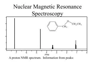
01/23/04. Biomolecular Nuclear Magnetic Resonance Spectroscopy. RESONANCE ASSIGNMENT IN PROTEINS Multi-dimensional NMR experiments NMR analysis of proteins. Proteins Have Too Many Signals! 1 H NMR Spectrum of Ubiquitin. ~500 resonances. Resolve resonances by multi-dimensional experiments.

E N D
01/23/04 Biomolecular Nuclear Magnetic Resonance Spectroscopy RESONANCE ASSIGNMENT IN PROTEINS • Multi-dimensional NMR experiments • NMR analysis of proteins
Proteins Have Too Many Signals! 1H NMR Spectrum of Ubiquitin ~500 resonances • Resolve resonances by multi-dimensional experiments
90º pulse Experiment (t) equilibration detection of signals Fourier Transform Data Analysis Time domain (t) The Pulse FT NMR Experiment
[2nd preparation] The 2D NMR Pulse Sequence + 1D + 1D = 2D
2D NMR: Coupling is the Key 2D detect signals twice (before/after coupling) 90º pulse t1 t2 Transfers between coupled spins Same as 1D experiment t1 t2
t1 t2 The 2D NMR Spectrum Pulse Sequence Spectrum Before mixing Coupled spins After mixing
The Power of 2D NMR:Resolving Overlapping Signals 1D 2 signals overlapped 2D 2 cross peaks resolved
Acronyms For Basic ExperimentsDiffer Only By The Nature Of Mixing Homonuclear Heteronuclear Scalar Coupling COSY HSQC TOCSY Hetero-TOCSY HMQC Multiple Quantum Dipolar Coupling NOESY NOESY-HSQC NOESY-HMQC
t2 t1 t3 Higher Dimensional NMR:Built on the 2D Principle 3D- detect signals 3 times 90º pulse (t3) Same as 1D experiment 3D NMR Pulse Sequence • Experiments are composites acronyms are composites
Proteins Have Too Many Signals! 1H NMR Spectrum of Ubiquitin ~500 resonances
Challenges For Determining Protein Structures Using NMR • Proteins have thousands of signals • Assign the specific signal for each atom • Thousands of interactions between atoms- also need to be assigned • Need to transform representation from spectrum through interactions between atoms to spatial coordinates
H H H H H H H H H H H NMR Spectrum to 3D Structure
Critical Features of Protein NMR Spectra • The nuclei are not all mutually coupled • Each amino acid gives rise to an independent NMR sub-spectrum, which is much simpler than the complete protein spectrum
Critical Features of Protein NMR Spectra • The nuclei are not all mutually coupled • Regions of the spectrum correspond to different parts of the amino acid • Tertiary structure leads to increased dispersion of resonances
Regions of the 1H NMR Spectrum What would the unfolded protein look like?
t2 t1 t3 Solutions to the Challenges • Increase dimensionality of spectra to better resolve signals: 123 4
Resolve Peaks By Multi-D NMR A BONUSregions in 2D spectra provide protein fingerprints If 2D cross peaks overlap go to 3D
Solutions to the Challenges • Increase dimensionality of spectra to better resolve signals: 123 4 • Detect signals from heteronuclei (13C,15N) • Better resolution of signals/chemical shifts not correlated between nuclei • More information to identify signals • Lower sensitivity to MW of protein
R R Double-Resonance ExperimentsIncreases Resolution/Information Content 15N-1H HSQC -15N - Ca- CO -15N - Ca H H
Large Scalar Couplings Less Sensitive to MW of the Protein • Superior to 1H homonuclear NMR: all JH-H < 20 Hz • Mixing is faster so less time for signal to relax