Download
1 / 43
470 likes | 815 Vues
HEMATOLOGIC MANIFESTATIONS OF SYSTEMIC ILLNESS. Prof. Mervat A Hesham 2010. hematologic manifestations mechanisms. 1. Bone marrow dysfunction a. Anemia or polycythemia b. Thrombocytopenia or thrombocytosis c. Leukopenia or leukocytosis

E N D
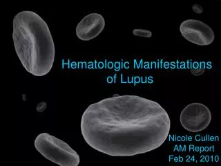
HEMATOLOGIC MANIFESTATIONS OF SYSTEMIC ILLNESS Prof. Mervat A Hesham 2010
hematologic manifestations mechanisms • 1. Bone marrow dysfunction a. Anemia or polycythemia b. Thrombocytopenia or thrombocytosis c. Leukopenia or leukocytosis • 2. Hemolysis • 3. Immune cytopenias • 4. Alterations in hemostasis a. Acquired inhibitors to coagulation factors b. Acquired von Willebrand disease c. Acquired platelet dysfunction • 5. Alterations in leukocyte function.
ANEMIA OFCHRONIC ILLNESS ● Normochromic, normocytic, occasionally microcytic ● Usually mild, characterized by decreased plasma iron. ● Impaired flow of iron from reticuloendothelial cells to the bone marrow ● Decreased sideroblasts in the bone marrow. Treatment involves treating the underlying illness. Iron is of little value because the iron is cleared by the reticuloendothelial system.
1 - Rheumatoid Arthritis ● Anemia of chronic illness (normocytic, normochromic) ● High incidence of iron deficiency ● Leukocytosis and neutropenia common in exacerbations of juvenile rheumatoid arthritis (JRA) ● Thrombocytosis , there may be transient episodes of thrombocytopenia.
2 - Systemic Lupus Erythematosus ● Two types of anemia are common: anemia of chronic illness (normocytic, normochromic) and acquired autoimmune hemolytic anemia (Coombs’ positive). ● Neutropenia is common as a result of decreased marrow production and immune mediated destruction. ● Lymphopenia with abnormalities of T-cell function. ● Immune thrombocytopenia. ● A circulating anticoagulant (antiphospholipid antibody) may be present and is associated with thrombosis.
3-Polyarteritis Nodosa ● Microangiopathic hemolytic anemia, possibly associated with renal disease or hypertensive crises ● Prominent eosinophilia 4- Felty’s Syndrome ● Triad of rheumatoid arthritis, splenomegaly, and neutropenia
5-Kawasaki Syndrome ● Mild normochromic, normocytic anemia with reticulocytopenia ● Leukocytosis with neutrophilia and toxic granulation of neutrophils and vacuoles ● Decreased T-suppressor cells ● High C3 levels ● Increased cytokines IL-1, IL-6, IL-8, interferon-á, and tumor necrosis factor (TNF) ● Marked thrombocytosis (mean platelet count of 700,000/mm3) ● DIC.
6-Henoch–Schönlein Purpura ● Anemia occasionally occurs as a result of GI bleeding or decreased RBC production caused by renal failure. ● Transient decreased F XIII activity may occur. ● Vitamin K deficiency from severe vasculitis-induced intestinal malabsorption.
7-Wegener Granulomatosis • This autoimmune disorder is rare in children. Hematologic features include: ● Anemia: normocytic; RBC fragmentation with microangiopathic hemolytic anemia ● Leukocytosis with neutrophilia ● Eosinophilia ● Thrombocytosis.
Infections A- Viral and Bacterial Illnesses Associated with Marked Hematologic Sequelae
A - Anemia OF INFECTIONS ●Chronic infection is associated with the anemia of chronic illness. ● Acute infection, particularly viral infection, can produce transient bone marrow aplasia or selective transient erythrocytopenia. ● ● Many viral and bacterial illnesses may be associated with hemolysis.
● Parvovirus B19 infection in people with an underlying hemolytic disorder (such as sickle cell disease, hereditary spherocytosis) can produce : - a rapid fall in hemoglobin - an erythroblastopenic crisis marked by anemia and reticulocytopenia. - There may be an associated neutropenia.
B- White Cell Alterations ● Viral infections can produce leukopenia and neutropenia. ●Neutrophilia with an increased band count and left shift frequently results from bacterial infection. ● Neonates, particularly premature infants, may not develop an increase in white cell count in response to infection. ● Eosinophilia may develop in response to parasitic infections.
C-Clotting Abnormalities • Severe infections can produce DIC. D - Thrombocytopenia • Infection can produce thrombocytopenia through decreased marrow production, • immune destruction, or DIC.
1- Parvovirus • transient erythroblastopenic crisis, particularly in individuals with an underlying hemolytic disorder. • thrombocytopenia, neutropenia, and a hemophagocytic syndrome. • In immunocompromised individuals, parvovirus B19 infection can produce prolonged aplasia.
2-Epstein–Barr Virus ● Atypical lymphocytosis ● Acquired immune hemolytic anemia ● Agranulocytosis ● Aplastic anemia ● Lymphadenopathy and splenomegaly ● Immune thrombocytopenia. ● immunologic and oncologic associations: - acquired hypogammaglobulinemia, and lymphoma - Clonal T-cell proliferations - Hemophagocytic syndrome - Endemic form of Burkitt’s lymphoma in Africa.
3- Human Immunodeficiency Virus • Thrombocytopenia occurs in about 40% of patients with AIDS Initially . • Thrombotic thrombocytopenic purpura (TTP) in advanced AIDS. • Anemia occurs in approximately 70–80% of patients and neutropenia in 50%. • Coagulation Abnormalities ● Dysregulation of immunoglobulin production may affect the coagulation cascade. ● Lupus-like anticoagulant (antiphospholipid antibodies) or anticardiolipin antibodies occur in 82% of patients. Thrombosis may occur secondary to protein S deficiency. Not due to Lupus-like anticoagulant .
Cancers in Children with HIV Infection • Non-Hodgkin lymphoma • Burkitt lymphoma (B-cell, small noncleaved) • Immunoblastic lymphoma (B-cell, large cell) • Central nervous system lymphomas • Mucosa-associated lymphoid tissue (MALT) type • Leiomyosarcoma and leiomyoma • Kaposi’s sarcoma • Leukemias
The pathogenesis of the hematologic disorders in HIV ●Infections:Myelosuppression is frequently caused by involvement of the bone marrow by infecting organisms (e.g., mycobacteria, cytomegalovirus [CMV],parvovirus, fungi, and, rarely, Pneumocystis carinii). ● Neoplasms:Non-Hodgkin lymphoma (NHL) in AIDS patients is associated with infiltration of the bone marrow in up to 30% of cases.
Medications:Widely used antiviral agents in AIDS patients are myelotoxic - zidovudine (AZT) causes anemia - Ganciclovir and trimethoprim/ sulfamethoxazole cause neutropenia. ● Nutrition:Poor intake is common accompanied by poor absorption. Vitamin B12 levels may be decreased due to : - vitamin B12 malabsorption - abnormalities in vitamin B12–binding proteins.
4- Torch Infections • neonatal anemia, jaundice. • thrombocytopenia, and HSM. 5- Bordetella Pertussis • marked lymphocytosis (>25,000/mm3) in early stages of infection. 6 - Tuberculosis • leukemoid reaction mimicking CML, monocytosis, and rarely pancytopenia.
7-Bartonellosis • Bartonella bacilliformis: fatal syndrome of severe hemolytic anemia with fever (Oroya fever). • Bartonella, B. henselae: - “cat scratch fever.” associated with a regional (following a scratch by a cat) lymphadenitis. - Thrombocytopenia may occur .
8-Leptospirosis (Weil Disease) This disease is caused by Leptospira icterohaemorrhagiae. • A coagulopathy occurs and can be corrected with vitamin K administration. • Thrombocytopenia commonly occurs but DIC is rare.
Infections B- Parasitic Illnesses Associated with Marked Hematologic Sequelae
1- Malaria Acute infections cause anemia which is multifactorial: ● Intracellular parasite metabolism alters negative charges on the RBC membrane,which causes altered permeability with increased osmotic fragility. ● Autoimmune hemolytic anemia may occur. An IgG antibody is formed against the parasite and resulting immune complex attaches nonspecifically to RBC, complement is activated, and cell destruction occurs. Positive Coombs’ test is found in 50% of patients. ● Thrombocytopenia without DIC is common.
2-Hookworm(Ancylostoma ) • Heavily infested children may present with - profound iron-deficiency anemia. - hypoproteinemia. - marked eosinophilia. 3- Leishmaniasis • splenomegaly and pancytopenia • The bone marrow usually is hypercellular with hemophagocytosis. • Some children may show coagulopathy.
4- Tapeworm(Diphyllobothrium latum) • worm infestation in the intestine results in vitamin B12 deficiency. 5- Trypanosomiasis • Adiagnosis of trypanosomiasis can be made by finding trypanosomes in a blood and bone marrow smear.
1- Protein-Calorie Malnutrition (kwashiorkor) • mild normochromic, normocytic anemia secondary to - reduced RBC production despite normal or increased erythropoietin levels. - reduced red cell survival. • impaired leukocyte function.
2-Scurvy • mild anemia is common. • bleeding tendency due to loss of vascular integrity, 3- Anorexia Nervosa • hypoplastic bone marrow, • Mild anemia (macrocytic), neutropenia, and thrombocytopenia. • Predisposition of infection associated with neutropenia
I. Nonneoplastic • A. Storage diseases 1. Gaucher disease 2. Niemann–Pick disease 3. Cystine storage disease • B. Marble bone disease (osteopetrosis) • C. Langerhans cell histiocytosis
II. Neoplastic • A. Primary 1. Leukemia • B. Secondary 1. Neuroblastoma 2. Non-Hodgkin lymphoma 3. Hodgkin lymphoma 4. Wilms’ tumor (rarely) 5. Retinoblastoma 6. Rhabdomyosarcoma
A- Nonneoplastic1- Gaucher Disease • Patients with Type 1 Gaucher disease present with: ● Hepatosplenomegaly (rarely, portal hypertension) ● Pancytopenia secondary to hypersplenism and rarely from infiltration of the bone marrow with Gaucher cells ● Bone pain, osteoporosis, pathologic fractures ● Growth delay ● Typical foamy cells in the bone marrow ● Erlenmeyer flask deformity of the distal femora on radiographs ● Decreased glucocerebrosidase activity of white cells ● Characteristic mutations of the glucocerebrosidase gene on chromosome 1 on DNA analysis.
2- Niemann–Pick Disease • The progressive deposition of sphingomyelin in the 1- central nervous system leads to type A, 2-in nonneuronal tissues leads to type B. 3- Type C is a neuropathic form that results from the defective cholesterol transport. • Niemann–Pick disease has classic signs, including: ● Hepatosplenomegaly ● Cherry red spot in macula ● Psychomotor deterioration ● Reticular pulmonary infiltrates ● Foamy cells in the bone marrow Diagnosis involves examining leukocytes or cultured fibroblasts to determine sphingomyelinase activity
3- Cystinosis ● Thermal instability, polydipsia, polyuria ● Failure to thrive ● Recurrent episodes of vomiting and dehydration ● Dwarfism and rickets often prominent ● Early renal involvement with tubular dysfunction manifesting as a secondary Fanconi syndrome, leading to chronic renal failure. Diagnosis ● Cystine crystals in the bone marrow ● Elevated cystine levels in leukocytes or fibroblasts.
4- Infantile Malignant Osteopetrosis (Marble Bone Disease) A- Severe Form (Autosomal Recessive) ● Progressive pancytopenia ● Compensatory extramedullary hematopoiesis with resultant leukoerythroblastic anemia (circulating normoblasts, tear-drop-shaped poikilocytosis, and early myelocytes), hepatosplenomegaly, and lymphadenopathy ● Bone marrow hypoplasia ● Hemolysis due to splenic sequestration of red cells and general overactivity of the reticuloendothelial system. B- Mild Form (Autosomal Dominant) Pathologic fractures occur in sclerotic bone. Nerve entrapment syndromes may also be present.
B- Neoplastic Disease ● Hemorrhage. ● Nutritional deficiency states. ● Dyserythropoietic anemias (including erythroid hypoplasia, sideroblastic anemia, and anemia similar to that seen in chronic inflammation). ● Defect in erythropoietin production. ● Hemodilution. ● Hemolysis. ● Pancytopenia secondary to marrow invasion or to cytotoxic therapy. ● Acquired von Willebrand disease as in Wilms’ tumor.
● Hypercoagulable states as in non-Hodgkin lymphoma. ● Coagulopathy as in acute promyelocytic leukemia. ● Leukoerythroblastic anemia. ● marrow Infiltration N.B : • Marrow infiltration is suspected when leukoerythroblastic anemia develops. This term signifies the presence of myelocytes and normoblasts with anemia, thrombocytopenia, and neutropenia. The explanation of this blood picture is that extramedullary erythropoiesis occurs when the marrow is infiltrated, permitting the escape of early myeloid and erythroid cells into the circulation. • Normal blood findings, however, do not exclude marrow infiltration.
“Foam Cells” in Bone Marrow “ 1. Neimann–Pick disease (types A, B, C, D) 2. Gaucher disease (types 1, 2, 3) 3. Gm1 gangliosidosis (type 1) 4. Gm2 gangliosidosis (Sandhoff variant) 5. Lactosyl ceramidosis 6. Sialidosis I 7. Sialidosis II, late infantile type 8. Mucolipidosis II 9. Mucolipidosis III 10. Mucolipidosis IV
11. Fucosidosis 12. Mannosidosis 13. Neuronal ceroid-lipofuscinosis 14. Farber disease 15. Wolman disease 16. Cholesteryl ester storage disease 17. Cerebrotendinous xanthomatosis 18. Chronic hyperlipidemia 19. Chronic corticosteroid therapy 20. Hematologic malignancies (e.g., Hodgkin disease, leukemia, myeloma) 21. Hematologic disease (e.g., aplastic anemia, ITP).