Download
1 / 16
160 likes | 253 Vues
Suggested HW Ch. 5. 1 – 9 (Chapter 5.1, 5.2) Note: Protein Explorer (originally due Friday) delayed. K d. What is the definition of K d ?

E N D
Suggested HW Ch. 5 • 1 – 9 (Chapter 5.1, 5.2) • Note: Protein Explorer (originally due Friday) delayed
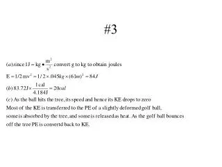
Kd • What is the definition of Kd? • Protein X interacts with Mg2+ with a Kd of 0.5mM. You have a solution of 0.1 mM Protein X. How much Mg2+ should you add so that the equilibrium concentration of complex (X•Mg2+) is 0.08 mM? • You do an experiment to measure the interaction between proteins Y and Z. When a solution of 0.15 pM Y and 0.56 nM Z reaches equilibrium, you determine that the concentration of Y•Z is 0.021 pM. What is the Kd describing the interaction?
Case study: oxygen binding in myoglobin and hemoglobin • Oxygen is poorly soluble in water (blood) • Iron (Fe2+)/O2 complex is soluble • But free iron is toxic • Use proteins containing an iron cofactor • Myoglobin • Hemoglobin
Iron is part of a heme prosthetic group: permanent association with protein “Porphyrin” ring
Iron has six coordination sites Four bind heme nitrogens One binds protein histidine “proximal” histidine His93/HisF8 One can bind O2
Structure of myoglobin • Extremely compact • “Globin” family • ~75% a helix (no b structure) • Eight helical segments • A-H • Four terminate in proline • Interior: hydrophobic except for two histidines
Proximal His coordinates Iron Distal His binds oxygen -increases affinity -decreases affinity for carbon monoxide CO still preferred over O2 -rotation (breathing) allows O2 exit & entry Distal His His64 His E7 Proximal His His93 His F8
“Globin” fold Six helices: “Three-over-three a-helical sandwich” Oxygen-carrying molecules Hemoglobins, myoglobins, cytoglobins, etc Heme-utilizing enzymes dehaloperoxidase Mammals Worms Fish Plants Bacteria
O2 binding by myoglobin • Reversible Myoglobin•O2 ↔ Myoglobin + O2 • O2 is a gas: use partial pressures (pO2) instead of molarity • Gas concentration proportional to pressure
Myoglobin: Hyperbolic dependence of O2 binding on pO2
Protein flexibility in myoglobin • Structural ‘breathing’ to allow O2 entry • Deoxymyoglobin vs. oxymyoglobin • Change in porphyrin ring, position of iron
Why hemoglobin (ie. why not just myoglobin)? • This is where the binding calculations get interesting • Oxygen carrier needs to ‘pick up’ O2 in oxygen-rich (pO2 > 10 kPa) blood surrounding lungs, & ‘drop off’ O2 in oxygen-poor tissues (pO2 ~ 4) • Hyperbolic binding of myoglobin: too insensitive to these types of Ds
Myoglobin: good at “picking up” O2, but won’t let go Tissues Lungs Little O2 “Dropped Off”
Hemoglobin: cooperative binding Much better O2 release