Download
1 / 29
320 likes | 532 Vues
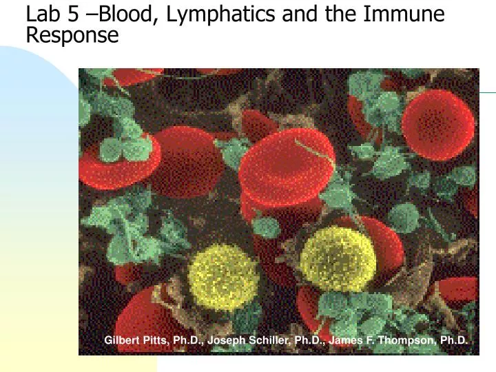
Lab 5 –Blood , Lymphatics and the Immune Response. Gilbert Pitts, Ph.D., Joseph Schiller, Ph.D., James F. Thompson, Ph.D. Objectives. Examine: Lymph node slide Lymphatic vessels on charts and models Blood slides

E N D
Lab 5 –Blood, Lymphatics and the Immune Response Gilbert Pitts, Ph.D., Joseph Schiller, Ph.D., James F. Thompson, Ph.D.
Objectives • Examine: • Lymph node slide • Lymphatic vessels on charts and models • Blood slides • Differentiate red blood cells, platelets, neutrophils, lymphocytes, monocytes, eosinophils, and basophils • Perform 2 differential WBC counts • Identify leukemia • Identify sickle cell anemia • Understand blood typing (ABO and Rh systems) • Calculate and interpret hematocrit/packed cell volume
The Lymphatic System • Basic organization • Lymph fluid in lymph vessels • Lymphatic organs (red bone marrow, thymus, spleen, lymph nodes, tonsils) with smaller collections of lymphatic tissue in other organs • Functions • Return interstitial fluid and proteins to the blood • Transport dietary fats to adipose tissue • Protect against cancer & infection • Lymph Flow from smallest to largest: • Capillaries vessels trunks ducts • Lymph vessels anastomose and supply and drain lymph nodes along their course
Lymph Flow Follows Venous Circulation • Right head, arm, and thorax drained by the right lymphatic duct into right subclavian vein • Left head, arm, thorax, most of the abdominal cavity and both legs drained by the thoracic duct into the left subclavian vein R L
The Lymph Node • Connective tissue capsule with trabeculaeextending from cortex to medulla • Stroma – the supportive connective tissue network of reticular fibers and fibroblasts trabeculae capsule
Circulation in the Lymph Nodes • Lymph enters via a number of afferent lymphatic vessels • It then enters a large subcapsular sinus and travels into a number of smaller sinuses • It meanders through these sinuses and exits the node at the hilus via efferent vessels • The node acts as a “settling tank,” because there are fewer efferent vessels, lymph stagnates somewhat in the node • This allows lymphocytes and macrophages time to carry out their protective functions Only lymph nodes filter lymph! Metastasis: cancer cells from a tumor break away and flow with the lymph until trapped in the lymph nodes
Lymph Node Parenchyma • Cortex - filled with lymphocytes and macrophages in follicles • Outer edge of follicle contains more T cells • Inner germinal center is the site of B-cell proliferation • Medulla - medullary cords of lymphocytes, macrophages, plasma cells (activated B cells) Cortex Medulla
Lymph Node Micrograph Medulla Cortex
Lymph Node Germinal Centers germinal centers
The Formed Elements of the Blood: • Leukocytes = White Blood Cells • Granular leukocytes (granulocytes) • neutrophils • eosinophils • basophils • Agranular leukocytes (agranulocytes) • lymphocytes - T cells, B cells • monocytes tissue macrophages
Eosinophil 2-4% Neutrophil 60-70% Basophil 0.5-1% Granular Leukocytes
Lymphocyte 20-25% Monocyte 3-8% Agranular Leukocytes
Leukocyte Life Span and Number • 5,000 - 10,000 WBC’s/mm3 blood • RBC/WBC ratio 700/1 • Differential WBC count (a standard clinical lab report) • Neutrophils 60-70% • Lymphocytes 20-25% • Monocytes 3-8% • Eosinophils 2-4% • Basophils 0.5-1% • Abnormal proportions are correlated with different types of disease processes
Eosinophil 2-4% Lymphocyte 20-25% Monocyte 3-8% Neutrophil 60-70% Basophil 0.5-1% Differential WBC Count
Leukocyte Identification Agranular Granular All have many large granules in cytoplasm & multilobed nuclei Dark Hidden nuc. Small Spherical nucleus Basophil Lymphocyte Red gran. Large 2+ lobes Eosinophil Faint gran. Monocyte no large granules in cytoplasm Neutrophil
Composition of Blood • Blood sample separates into 2 parts • plasma - straw colored liquid on top • ~55% of the volume • formed elements • ~45% of the volume • red blood cells • buffy coat: white blood cells and platelets
Hematocrit (Hct) • Packed Cell Volume is the % of the blood which is RBC’s • Males: 40-54% (47%) • Females: 38-46% (42%) • Hct indicates the status of RBC production, the state of hydration, or various disease states
Blood Typing • Antigen – any substance which provokes specific immune responses • Antigenic determinants • Antigen parts which trigger the specific immune response • An antigen may be an entire microbe or only small structures such as subregions of large molecules • RBC antigens (agglutinogens) are membrane glycoproteins Most “antigens” are complex and express multiple types of antigenic determinants.
ABO Blood Types • 2 glycoprotein agglutinogens, A & B • One gene from each parent, A, B or O • 6 combinations - AA, AB, AO, BB, BO, OO (no agglutinogens)
ABO Blood Types • Agglutinins • Naturally occurring antibodies produced in response to the agglutinogens not present in your blood • React in antigen-antibody response to blood not of your type • blood type AB = universal recipients • blood type O = universal donors
Rh System • Rh typing - Rhesus monkey • Those expressing Rh antigens are Rh+ • Those without Rh agglutinogens are Rh- • Normally, blood does not contain Rh agglutinins • Immune system only makes agglutinins in response to specific exposure to Rh antigens • Rh sensitivity does not occur until second transfusion • Hemolytic disease of the newborn (erythroblastosis fetalis) • many “blue babies” prior to WWII
Please Clean Up Your Work Area • Place only lancet and capillary tubes in designated sharps containers • Place all other blood contaminated materials (gloves, alcohol wipes, paper towels, etc.) in the large red biohazard bucket at the front of the room • Place all other discards in regular trash receptacles • Disinfect your work area with the spray solution after you have concluded your blood work.
Homework • Complete and turn in the questions on pages 5-13 to 5-15 • Complete Assignment 5 on MasteringAandP.