Download
1 / 1
10 likes | 65 Vues
Reproducibility of Functional MRI – Progress Towards Profile Development.

E N D
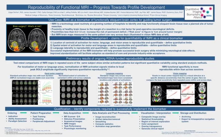
Reproducibility of Functional MRI – Progress Towards Profile Development Edgar DeYoe1, PhD, James Voyvodic2, PhD,Cathy Elsinger, PhD (co-chair)3, Jeffrey Petrella, MD (co-chair)2, Harris Ahmad, MD4, Andrew Buckler, MS5, Feroze Mohamed, PhD6, Jay Pillai, MD7, James Reuss, PhD8, Domenico Zaca, PhD7 1Medical College of Wisconsin,2Duke University, 3NordicNeuroLab,4BioClinica, Inc, 5Buckler Biomedical, 6Temple University, 7John’s Hopkins University Medical School, 8Prism Clinical Imaging Use Case: fMRI as a biomarker of functionally eloquent brain cortex for guiding tumor surgery • fMRI is a technology used routinely at a growing number of hospitals to identify and map functionally eloquent brain tissue near a planned site of tumor resection. • Proximity of healthy brain tissue to the margin of a resection is a risk factor for post-operative neurological deficits. • Proximities less than 0.5-1.0 cm increase the risk of permanent deficit. (“Risk zone” in figure is 1cm around tumor margin) • But fMRI brain maps measured in the same patient can vary across days (illustrated in vision fMRI data at left). Profile Development – claims for quantifiability of fMRI as a clinical biomarker 1) Location of centroid of activation for motor, language, and vision areas is reproducible and quantifiable – define quantitative limits 2) Spatial extent of activation for motor and language areas is reproducible and quantifiable – define quantitative limits 3) Language laterality is reproducible and quantifiable– define quantitative limits Successful implementation of the fMRI biomarker will help promote successful brain surgery while minimizing neurological side-effects. Improved standardization will facilitate adoption as standard of care and promote industry-wide acceptance. Preliminary results of ongoing RSNA-funded reproducibility studies Test-retest comparisons of fMRI maps in repeated scans of the same subject show similar activation patterns but significant quantitative variability using standard analysis methods. For localization of motor or language areas, normalization of fMRI activation signals relative to local fMRI functional specificity is more peak BOLD amplitude significantly improves quantitative reproducibility1,2. reproducible within than across sessions Hand motor mappingLanguage mappingVision mapping Standard activation maps vary with scan duration Normalized extent of activation is Standard t-maps vary across scans Voxels in visual cortex respond best to a visual stimulus at a AMPLE normalized maps are stable over scan time consistent within and across subjects Normalized maps have more consistent specific distance (eccentricity) from the center of gaze. Here we locations and extents of activations compare the best eccentricity from repeated scans within and across days. 6 scans of one subject 2 scans for each of 6 subjects Qualification – Identify components required to successfully implement the biomarker Ordering Patient Preparation Data Acquisition Data Analysis and Post Processing Visualization / Interpretation Storage and Distribution • Indication • Ability Assessment • Paradigm Selection • Task Training • Positioning • Peripherals Adjustment • MR Scanner Q/A • Stimulus Presentation • Anatomical Images • fMRI images • Task Performance • Physiologic monitoring • Archiving • Export to intraoperative navigation system • Composite image overlay • Statistical thresholding • Region of interest exploration • Color assignment • Overlay transparency • Generate clinical report • Image reconstruction • Artifact detection/removal • Smoothing • Compute statistics • Activation map generation References 1. J.T. Voyvodic (2006) Mag. Res. Imag., 24:1249-1261. 2. J.T. Voyvodic, J.R. Petrella, and A.H. Friedman (2009) J. Mag. Res. Imag. 29:751-759. 3. http://qibawiki.rsna.org 4. http://www.fda.gov/downloads/Drugs/GuidanceComplianceRegulatoryInformation/Guidances/UCM230597.pdf Acknowledgements The members of the QIBA fMRI Technical Committee would like to acknowledge the continued support of the RSNA and staff devoted to support of the QIBA activities.