Download
1 / 16
160 likes | 346 Vues
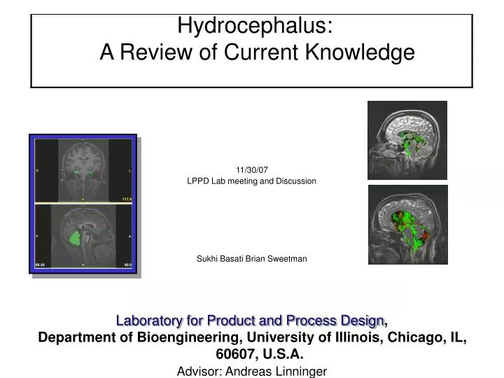
Hydrocephalus: A Review of Current Knowledge. 11/30/07 LPPD Lab meeting and Discussion Sukhi Basati Brian Sweetman Laboratory for Product and Process Design , Department of Bioengineering, University of Illinois, Chicago, IL, 60607, U.S.A. Advisor: Andreas Linninger.

E N D
Hydrocephalus: A Review of Current Knowledge 11/30/07 LPPD Lab meeting and Discussion Sukhi Basati Brian Sweetman Laboratory for Product and Process Design, Department of Bioengineering, University of Illinois, Chicago, IL, 60607, U.S.A. Advisor: Andreas Linninger
Dandy, 1919 Occluded various veins, foramen, aqueduct and observed development of HC. Also replaced CSF with ink and observed flow of CSF into different regions. Showed SAS is primary site of absorption. Method: Used cotton swabs in dogs for occlusions. Implanted gauze for CH Findings: lateral and third ventricles enlarged but 4th did not with occusion of aqueduct. Obliterate both lateral ventricles by occluding both foramina of monro and obstruct the aqueduct and 3rd ventricle enlarges. Meanings: says communicating HC is an overproduction of CSF. In communicating HC, SAS reduction in area of 1/5.
Dandy, 1919 Experiment a) Implanted cotton swabs into aqueduct of sylvius. Did not allow enlargement of the head by surgical sutures. Results lateral and third ventricules enlarged but 4th did not. Experiment b) Occluded foramina of monro with small piece of fascia (connective tissue). Result saw no change in other ventricle. Experiment c) Removed choroid plexus and blocked foramen Result saw ventricle become small. Experiment d) Blocked both sides of foramen and kept 1 choroid plexus intact Result ventricle collapse without choroid plexus.
Dandy, 1919 Experiment e) Obstructed large vein of galen and produced HC Result HC develops when an occlusion of the great vein of Galen is located at its origin (a specific location in the vein) Experiment f) Replaced CSF with ink in dogs and killed at various stages. Result cisternae fills rapidly along with cerebellar SAS. Sulci of hemispheres fills slowly. Experiment g) Produced adhesions around midbrain with gauze and irritant and injected ink to observe flow. Result no ink flowed into SAS.
Berring, 1955 Method: Used dogs and patients undergoing examination. Occluded foramen via gelatin sponges soaked in kaolin. Recorded pressures via manometers. Recorded pressure from cerebral ventricle, carotid artery, right atrium. Also compared ECG. Findings: Due to phase lags of pressure waves, claims that a pressure gradient exists throughout CSF system which sets up fluid flow. CSF pulse is generated intracranially by the arterial pulsation of the blood and not through local venous pressure changes. Also determines contribution of CSF to mean ICP. Meaning: decrease in magnitude of pulse may be caused by: a)damping by elastic dural sac b)small changes in craniospinal blood volume. c)Escape of small amounts of csf into SAS. CSF pulse gets bigger with increase in ICP d) minor fluctuations of CSF volume can be easily absorbed by an equal change of one or a combination of volume controlling variables. (as HC develops, space is confined, so pulse cannot be damped) Induced plexoctomy and occlusion of foramen of monro to determine pulse pressure waves.
Mean ICP (55 dogs) 99 mm H20 (45 – 145 mm H20) (18 = std. Dev) After choroids plexectomy 72 mm H20 Berring, 1955 Experiment a) Unilateral plexectomy and plug foramen of monro in 6 dogs. Control = unilateral plex. Without occlusion of foramen. Results: remove choroid plexus and plug foramen = no pulse But you get normal pulse in opposite ventricle. Dog with plex. And open foramen = decreased pulse. Experiment b) Determine the contribution of CSF to mean ICP. Method: measure cisternal CSF pressure before and after removal of choroid plexus of 22 dogs. Control: ventriculotomy without plex. = 5 dogs. Note: it was common to do choroids plexectomy on adults in those days without ill effects.
Berring, 1955 Experiment d) Intraventricular pulse pressure increases with body length(age) Recorded from many patients. Increase in choroids plexus = increase in intraventricular pulse pressure But no variation in CSF production or flow Suggests reason of CSF flow being development of embryonic subarachnoid pathways.
Di Rocco, 1978 Communicating hydrocephalus induced by mechanically increased amplitude of the intraventricular cerebrospinal fluid pulse pressure: Rationale and Method Summary: 1. Balloons were inserted into the lateral ventricles of lambs. 2. The increased ventricular CSF pulsations from the pulsating balloon caused communicating hydrocephalus in all animals, although the mean ventricular CSF pressure was normal. 3. The experiment was inspired by the idea that increased ventricular CSF pulsations could cause communicating hydrocephalus and by Bering’s suggestion that increased pulsations in the choroid plexus could act as a “water hammer” pulse within the ventricles. 4. This is the first experimental proof that increased CSF pulse pressure can cause hydrocephalus and that ventricular enlargement may develop without CSF obstruction and without increase of mean CSF pressure.
Shows how a delay in the applied pulse pressure affects the ICP wave. Maximal constructive interference=delay of 60 to 100ms from the R wave in the ECG Shows that different input waves have different affects on the mean ICP. The duration of A’ is longer than B’ and requires fluid to be extracted from the balloon to maintain normal ICP values
Major Conclusions • 1.The increase in the amplitude of the CSF pulse pressure, even if induced for a relatively short period (3 h) is followed by significant neuropathic lesions • 2. The characteristics of the lesion, their extent, as well as their location are directly related to the amplitude of the pulse pressure (3x vs 6x the control value) and to the site where the source for mechanical pulses is inserted • 3. For provoking a ventricular dilation the pulses must originate from within the ventricles.
Del Bigio/Bruni, 1987 Experiment: Specific gravity measurements made on fresh and dried samples of hydrocephalic and normal rabbit brains. Method: Control group injected w/ NaCl; Test group injected w/ silicone oil into the cisterna magna. Coronal slices were used to determine/confirm ventricular enlargement. Cylindrical slices (0.5mm thick) from the cortical surface to the level of the ventricle analyzed. Cerebral water content in silicone oil-induced hydrocephalic rabbits
Findings: • Density gradient found in normal cortical gray matter (density increased in the direction of C1 to M, figure above, left). • Data obtained from control rabbits indicate that superficial cortical gray matter has a lower specific gravity (higher water content) than white matter (conceptual slide below) • Gray matter from deeper cortical layers has a higher specific gravity (less water content) than white matter. • Hydrocephalus accompanied by significantly increased specific gravity (decrease in water content) at 3 days and 1 and 4 weeks. • In the 1 and 4 weeks groups, there were significant increases in the specific gravity of white matter in the corpus callosum (V2). • Loss of extracellular brain water appears to compensate for the enlarging cerebral ventricles • No changes in the composition of the anhydrous cerebral tissues of the hydrocephalic rabbits • Because the constituents of solid cerebral tissue were unchanged, the specific gravity changes in fresh brain are attributable to decreased water content
Meaning: • These changes represent water loss throughout the whole cerebrum except at the ventricular surface (where little change in specific density was observed). The specific gravity of solid tissue brain components changed insignificantly (figure above, right). • Short-term brain volume changes in experimental hydrocephalus were due mainly to loss of tissue water. Conceptual only Experiment was done on rabbits Normal: Relative water content within in the parenchyma. 10=highest level; 5 the lowest Hydrocephalic: Relative water content within in the parenchyma. 10=highest level; 5 the lowest