Download
1 / 1
10 likes | 99 Vues
Results / Further work . Abstract.

E N D
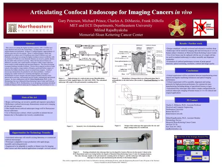
Results / Further work Abstract Skin cancers are among the highest incidence cancers, with 1.2 million new cases detected each year in the United States. To detect these new cases, 5.5 million biopsies are performed. Of these 5.5 million biopsies, approximately 4.3 million cases turn out to be normal. These 4.3 million biopsies, which may potentially be avoided, costs US healthcare more than $2 billion every year. Similarly, oral cancers are among the highest incidence cancers worldwide, with an estimated 274,000 new cases detected. Biopsies are invasive, painful, destroy the site under study and leaves scarring. Often, the best areas to biopsy are difficult to ascertain, and a small number of biopsies within a large suspicious lesion results in under-sampling and uncertain diagnoses. The tissue processing to prepare histology introduces artifacts. Precise micro-surgical excision of large cancers often requires a large number of biopsies, performed before surgery, to determine the hidden subsurface cancer-to-normal tissue margins. Confocal microscopy demonstrates the potential to solve these problems. Confocal imaging may enable noninvasive screening and diagnosis, pre-surgical determination of cancer margins and intra-surgical guidance directly on the patient and in real-time, with minimal need for biopsy, minimal pain and minimal expense. In close collaboration with Dr. Milind Rajadhyaksha, Ph.D., in the Dermatology Service at Memorial Sloan-Kettering Cancer Center and Professor Charles DiMarzio, Ph.D. (Northeastern), we are designing a prototype articulated toothbrush-shaped confocal endoscope for imaging skin and oral tissues in vivo. • Design completed, critically reviewed and released to machine shop • Endoscope will be tested for imaging skin and oral mucosa in vivo on human subject volunteers at Memorial Sloan-Kettering Cancer Center • Use of two lenses may be replaced with the use of a single rod lens (as used in standard endoscopy) to reduce assembly and alignment constraints • Evaluation of confocal performance in terms of point spread functions, optical sectioning, resolution, contrast and image quality Introduction Enter text and box will expand State of the Art • Biopsy and histology are invasive, painful and expensive procedures • Reflectance confocal microscopy demonstrates noninvasive imaging of skin and oral cancers • Fiber bundle-based confocal endoscopes image well in fluorescence but not reflectance • Use of fluorescence contrast, which is possible in animals but not humans due to fluorophore dye toxicity considerations Opportunities for Technology Transfer • Articulated endoscope will retrofit existing laboratory or commercial confocal microscopes • Prototype is adaptable to mass production with rapid design, assembly and testing protocols • Engineered to be adaptable to smaller or thinner sizes for imaging other tissues and other medical applications (example: cervical cancer) Conclusions PI Contact • Critical requirement will be correlation between manufacturing errors (lateral and angular machining tolerances) and optical imaging performance • Critical requirement will be assembly, alignment and testing protocols for high-precision performance for commercial mass-manufacturing (including design of specialized fixtures or jigs). • Articulated relay telescopes offer relative simple configurations for confocal endoscopic imaging of human tissues in vivo for clinical and surgical applications Charles A. DiMarzio, Ph.D., Associate Professor Electrical and Computer Engineering Northeastern University Boston MA 02115 dimarzio@ece.neu.edu . Milind Rajadhyaksha, Ph.D., Assistant Member Dermatology Service Memorial Sloan-Kettering Cancer Center 160 East 53rd St. New York NY 10022 rajadhym@mskcc.org Articulating Confocal Endoscope for Imaging Cancers in vivo Gary Peterson, Michael Prince, Charles A. DiMarzio, Frank DiBella MET and ECE Departments, Northeastern University Milind Rajadhyaksha Memorial-Sloan Kettering Cancer Center Figure 1. Optical design of a confocal microscope [Rajadhyaksha, Anderson & Webb, Applied Optics 38: 2105-2115, 1999]. The articulated endoscope will couple at the galvanometric scanning mirror. Figure 2. Morphology of human skin in an orthogonal plane that is perpendicular to the skin surface [Borysenko L, Functional Histology (p. 122), 1979]. Figure 4. Design of the endoscope that approaches the size and shape-configuration of a toothbrush. Figure 3. Isometric view of articulating endoscope. Figure 5. Existing articulated relay telescope that was developed by Gustavo Herrera for his master’s thesis in the ECE Department and is now being further developed at Memorial Sloan-Kettering Cancer Center for imaging humans. The relay telescope couples at the galvanometric scanning mirror in Figure 1, and the endoscope will couple to this telescope to create an opto-mechanical probe interface to the human subject. This work is supported in part by the Center for Subsurface Sensing and Imaging Systems, under the Engineering Research Centers Program of the National Science Foundation (Award Number EEC-9986821).