Download
1 / 17
170 likes | 318 Vues

E N D
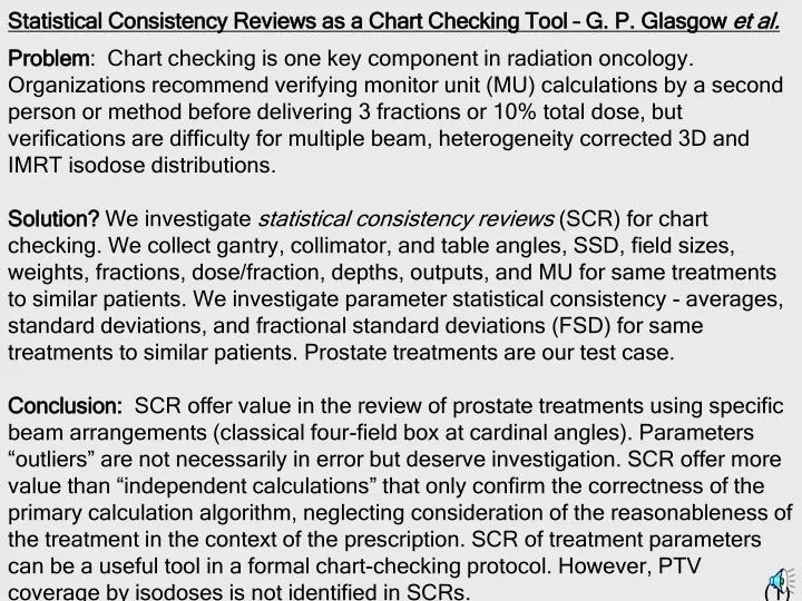
Problem: Chart checking is one key component in radiation oncology. Organizations recommend verifying monitor unit (MU) calculations by a second person or method before delivering 3 fractions or 10% total dose, but verifications are difficulty for multiple beam, heterogeneity corrected 3D and IMRT isodose distributions. Solution? We investigate statistical consistency reviews (SCR) for chart checking. We collect gantry, collimator, and table angles, SSD, field sizes, weights, fractions, dose/fraction, depths, outputs, and MU for same treatments to similar patients. We investigate parameter statistical consistency - averages, standard deviations, and fractional standard deviations (FSD) for same treatments to similar patients. Prostate treatments are our test case. Conclusion: SCR offer value in the review of prostate treatments using specific beam arrangements (classical four-field box at cardinal angles). Parameters “outliers” are not necessarily in error but deserve investigation. SCR offer more value than “independent calculations” that only confirm the correctness of the primary calculation algorithm, neglecting consideration of the reasonableness of the treatment in the context of the prescription. SCR of treatment parameters can be a useful tool in a formal chart-checking protocol. However, PTV coverage by isodoses is not identified in SCRs. (1) Statistical Consistency Reviews as a Chart Checking Tool – G. P. Glasgow et al.
Recommendations Regarding Monitor Unit Calculations & Chart Checks 1994 - AAPM TG40, Comprehensive QA...Oncology - that graphical isodoses ...be checked by “…an independent calculation of dose at one point in the plan...at the isocenter or a point near the center of the tumor”; “For no graphical plans “…monitor unit check…reviewed by another ... individual...a radiation oncology physicist…before the third fraction or before 10% of the dose has been delivered....”1 1998- AAPM TG53, QA for...Planning... - “…standard clinical protocol cases should be planned, and MUs calculated for each field.”“The doses actually deliverer ... verified independently ...by measurement or ...standard MU calculation data” It recommends verifying monitor unit (MU) calculations by a second person or method before delivering 3 fractions or 10% dose. 1997 – ACR Guidelines .. 3-D & Conformal Therapy states, regarding software verification... QMP should “Confirm .. accuracy .. calculated MUs.”3 2002 - ACR Practice Guidelines for IMRT notes, “As patient data are accumulated that demonstrate dosimetric accuracy of the IMRT planning/delivery system, dose and dose distributions alternately may be verified using an independent dose calculation method. 4 (2)
2002-NRC 10CFR Part 35, Section 35.41 requires licensees “...develop, implement...written procedures...checking...manual and computer ...dose calculations.”8 2003 -AAPM Report 80- Solo Practice of Medical Physics... includes “Verifying ... dosimetry calculations were checked by a second person or method, before the lesser of three fractions or 20% of the dose delivered. 5 Note the increase to 20% of dose delivered from the earlier 10% dose delivered. 2 2004 - ACR Technical Standards ... External Beam Therapy amends its earlier recommendation, dropping the 10% dose delivery rule; stating “All… MU calculation…shall by verified by another person or method before the first treatment if the total number of fractions is five or fewer or otherwise before the third fraction.” 6 Medical physicists “…shall develop a chart check protocol…” and states “A physics chart review shall be conducted weekly.” 6 2004 ACRO Guidelines …Practice Accreditation Program states “…calculations must be independently checked by another person or another method of calculation ... before administration of the third fraction and at any time that any changes are made.”7 (3)
Conclusion 1: MU calculations need to by timely checked, preferable by a second individual, using either an “independent methods” or by “other methods” in a manner sufficient to prevent irreparable harm to the patient from incorrect MU prescriptions. Methods of confirming “checks” are not prescribed in reports. Conclusion 2: Current MU verification is time-consuming and difficulty for multiple beams, heterogeneity corrected 3D and IMRT isodose plans.Current treatments involve more than 20 arbitrary shaped fields, at non-cardinal angles, and use heterogeneity corrections. Alternative confirmation calculations constitute an onerous QA requirement. Conclusion 3: Commercial software addresses some needs of confirmation calculations. But, as noted in AAPM TG-53, some independent methods may lack the accuracy of the MU calculations from the isodose computer.2 We investigate statistical consistency reviews (SCR) potentiallyapplicable to common treatments, as one chart-checking tool. For some treatments, reviewing MU calculations for statistical consistency againsta database of confirmed MUs may be a reasonable substitute for more time-consuming MU verification methods. SCR is not applicable for all treatments nor all MU verifications. We report initial results for prostate treatments(4)
Data for 22 post-surgery patients – Single PTV; 66.6 Gy; 1.8 Gy; 37 Fxs
Data for 13 low-risk patients- Single PTV; 74 Gy; 2.0 Gy; 37 Fxs
Datafor 14 intermediate-risk patients – Two PTVs; 75.6 Gy; 1.8 Gy; 42 Fxs
Data for 12 high-risk patients- 3 PTVs; 75.6 Gy, 1.8 Gy; 42 Fxs
ANALYSIS Classical 4-field cardinal angle (0o, 90o, 180o, 27 0o) prostate treatments for all patients had similar fractional standard deviations (FSD) for SSD (2%), collimator eq.sq. field size (5%-10%), and effective treatment depths (8%-17%). Single PTV patients had consistent FSD beam weight (2%-4%) & MU (4%-6%). Patients’ PTV2 exhibited the largest FSD for beam weight (15-30%) and MU (4%-8%), reflecting differing MDs decisions on the exact “boost” dose given to PTV2. Five-field (0, 75, 140, 220, 285 degree) IMRT treatments beam weight and MU exhibited large FSD (14% - 21%). They are smaller than expected; they still can be used to flag, for investigation, unusually small or large beam weights or MU in IMRT treatments. However, the nature of IMRT calculations makes it less likely than a parameter unusually smaller or larger than the normal parameters may be in error. There are only 16 patients in this data set. (12)
How can we use these data? Rather than check our known correct algorithm MU calculations against another algorithm, always agreeing, we review prostate MU calculations against statistical averages, focusing not on the accuracy of the algorithm calculations but on the likely correctness of the treatment prescription. We quickly check parameters visually against a review table. A recent patient’s AP 57 MUs were more than 3 FSD from the average 47 +/- 2.5 MU. His AP 79 cm SSD was below the average 87.9 +/- 1.9 cm. Our review flagged the case. A planning error? No, the patient was just obese; but it illustrates knowing average treatment parameters for the prostate. Isodose distribution deviations, e.g. inadequate PTV coverage, cannot be revealed by SCR. A PTV treated with inadequate field sizes yielding inadequate PTV coverage may appear acceptable as treatment parameters may still be near the averages. Chart checks require reviews of printouts of the visual parameters, e.g., multileaf collimator placements relative to PTVs and review of dose-volume histograms to confirm that 95% of the prescribed absorbed dose covers 95% of the PTV, or similar criteria. (13)
How can we use these data? Rather than check our known correct algorithm MU calculations against another algorithm, always agreeing, we review prostate MU calculations against statistical averages, focusing not on the accuracy of the algorithm calculations but on the likely correctness of the treatment prescription. We quickly check parameters visually against a review table. A recent patient’s AP 57 MUs were more than 3 FSD from the average 47 +/- 2.5 MU. His AP 79 cm SSD was below the average 87.9 +/- 1.9 cm. Our review flagged the case. A planning error? No, the patient was just obese; but it illustrates knowing average treatment parameters for the prostate. Isodose distribution deviations, e.g. inadequate PTV coverage, cannot be revealed by SCR. A PTV treated with inadequate field sizes yielding inadequate PTV coverage may appear acceptable as treatment parameters may still be near the averages. Chart checks require reviews of printouts of the visual parameters, e.g., multileaf collimator placements relative to PTVs and review of dose-volume histograms to confirm that 95% of the prescribed absorbed dose covers 95% of the PTV, or similar criteria. (13)
Conculsions: For highly systematic treatments, such as the prostate, reviewing MU calculations for statistical consistency againsta database of prior confirmed MU may be a reasonable substitute for more time-consuming MU verifications. A quick review of MU calculation parameters allows one to ascertain the reasonable correctness of the calculation and also the proposed treatment. An outlier parameter is not necessarily in error but signals the need for investigation. All anatomic sites and treatment methods are not amenable tostatistical consistency reviews nor are we recommending it for all MU verifications. However, initial results for prostate treatments are encouraging; we are studying other sites and treatments to which the method may apply. The results of SCR are an excellent learning tool for those in training. (15)
Abbreviated References without Authorship 1. AAPM TG-40 Comprehensive QA ... oncology: Med Phys.21: 581 – 618; 1994. 2. AAPM TG53: Quality Assurance ... planning. Med Phys.25: 1773 – 1829; 1998. 5. AAPM Report 80. Solo Practice... Oncology. Madison, WS: Med Physics Pub; (2003.) From Practice Guidelines & Technical Standards 2005. Reston, VA: ACR 2005 3. ACR ... Guideline for 3-D External Beam ... Planning and Conformal Therapy. (1997). 4. ACR Practice Guideline for Intensity-Modulated Radiation Therapy. (2002). 6. ACR Technical Standard...Radiation ... Physics for External Beam Therapy (2004). 7. ACRO Red Book; Guidelines... Practice Accreditation Program. Toledo, OH; (2004). 8. NRC. 10 CFR 35 (Medical use of byproduct material: final rule.) [Online]. (16)