Download
1 / 59
610 likes | 934 Vues
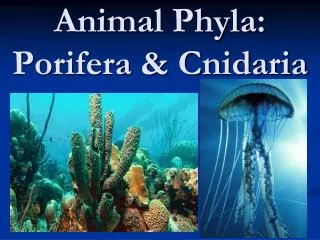
Phyla Porifera, Cnidaria, and Ctenophora. Chapter 9. Phylum Porifera (Sponges). Poriferans are mostly marine animals. Characteristics include: Asymmetrical or radial symmetry Three types of cells Central cavity for water circulation No tissue or organs Mostly sessile.

E N D
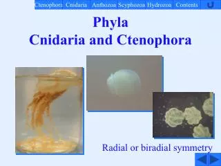
Phyla Porifera, Cnidaria, and Ctenophora Chapter 9
Phylum Porifera (Sponges) • Poriferans are mostly marine animals. • Characteristics include: • Asymmetrical or radial symmetry • Three types of cells • Central cavity for water circulation • No tissue or organs • Mostly sessile
Poriferan Cell Types • Pinacocytes • Thin, flat cells that line the outer surface of sponges • Some specialized into porocytes that regulate water circulation • Amoeboid cells • Contained in jellylike mesohyl layer • Function in reproduction, secreting skeletal elements, transporting and storing food, and forming contractile rings around openings in the wall • Choanocytes • Collar cells lining inner chamber • Flagellated with microvilli surrounding each flagellum • Flagella create water current and the collar of microvilli filters food particles
http://www.waycross.edu/faculty/gcook/ecology/animalia/zoa.htmlhttp://www.waycross.edu/faculty/gcook/ecology/animalia/zoa.html
Sponge Skeleton • Sponge skeletons may be made of needlelike spikes called spicules. • Spicules are made of calcium carbonate or silica and are formed by amoeboid cells. • Some sponges have skeletons made of spongin which is made of collagen • Spongin skeletons are used as commercial sponges
http://www-biol.paisley.ac.uk/biomedia/graphics/jpegs/SPICULES.gifhttp://www-biol.paisley.ac.uk/biomedia/graphics/jpegs/SPICULES.gif
Ascon Body Form • Vaselike shape • Outer openings of porocytes are called ostia (singular-ostium) • Ostia lead directly to inner chamber called the spongocoel which is lined with choanocytes • Water is drawn in through ostia and exits through large opening at the top of the sponge called the osculum.
http://mac01.eps.pitt.edu/geoweb/courses/GEO1200/lab3/structure.htmhttp://mac01.eps.pitt.edu/geoweb/courses/GEO1200/lab3/structure.htm
Sycon Body Form • Outer wall appears folded • Water enters through openings called dermal pores which are openings of incurrent canals (infolded invaginations). • Incurrent canals are connected to radial canals through pores. • Radial canals are lined with choanocytes and lead to the spongocoel. • Spongocoel has an osculum.
http://mac01.eps.pitt.edu/geoweb/courses/GEO1200/lab3/structure.htmhttp://mac01.eps.pitt.edu/geoweb/courses/GEO1200/lab3/structure.htm
Leucon Body Form • Leucon body forms contain an extensively branched canal system. • Water enters through ostia and enters incurrent canals. • Incurrent canals lead to choanocyte chambers and leaves chambers through excurrent canals. • No spongocoel is present and there are multiple oscula.
http://mac01.eps.pitt.edu/geoweb/courses/GEO1200/lab3/structure.htmhttp://mac01.eps.pitt.edu/geoweb/courses/GEO1200/lab3/structure.htm
Nutrition and Gas Exchange • Sponges feed on bacteria, microscopic algae, protists, and other microscopic organisms. • Some deep water sponges (Genus Asbestopluma) are carnivorous and feed on small crustaceans. • Sponges serve the ecosystem by filtering water and reducing turbity (cloudiness). • A 1 cm by 10 cm sponge can filter 20 liters of water every day. • Food is filtered and trapped by choanocyte cells and then trapped by food vacuoles where digestion by enzymes begins. • Amoeboid cells distribute digested food products to other cells. • Nutrients can also be phagocytized by pinacocytes along incurrent canals or absorbed by active transport. • Gas exchange and excretion of nitrogenous wastes occur by diffusion
Asbestopluma http://scilib.ucsd.edu/sio/nsf/fguide/porifera26.html
Cellular Communication • Sponges do not have a nervous system for cell to cell communication • Cells respond to stimuli in the environment to regulate activities. • Example: Sunlight inhibits constriction of ostia keeping them open, thus maintaining maximum water circulation during sunlight hours. • Some cellular communication may be present due to observations of activity changes with no external stimulus. • Method of communication is unknown.
Reproduction • Most sponges are monoecious meaning both sexes occur in one organism. • Egg and sperm are produced at different time to prevent self fertilization. • Egg and sperm are derived from meiotic choanocytes. • Eggs are stored in mesohyl. • Sperm exit one sponge through its osculum and enter another with incurrent water. • Sperm are transferred to egg by amoeboid choanocytes. • Asexual reproduction occurs in some marine sponges and freshwater sponges. • Gemmules containing amoeboid cells are released during winter when the parent dies. The gemmules release the amoeboid cells during the spring which organize into a sponge.
Life Cycle of Sponges • Earliest development occurs in the mesohyl. • The zygote cleaves and forms a flagellated larva. • Larva break free of the mesohyl and exit the sponge through water currents. • The larva free swims for 2 days and finally settles on a substrate and develops into an adult sponge.
Porifera Classification • Class Calcarea • Calcium carbonate spicule skeletons • All three body forms represented • All marine • Also known as calcareous sponges • Examples: • Grantia • Leucosolenia
http://www.bscd.uchicago.edu/classes/biosci184/Images/Grantia.htmlhttp://www.bscd.uchicago.edu/classes/biosci184/Images/Grantia.html http://www.dscc.edu/kjones/bio2animal.htm http://www.olympusmicro.com/micd/galleries/darkfield/grantia.html
http://www.mareco.org/KML/sponges/pages/leucosolenia%20species_jpg.htmhttp://www.mareco.org/KML/sponges/pages/leucosolenia%20species_jpg.htm http://www.mareco.org/KML/sponges/pages/leucosolenia%20eleanor_jpg.htm http://www.biol.rug.nl/onderwaterbiologie/foto10.jpg
Classification (cont.) • Class Hexactinellida • Silica spicule skeletons • Spicules often form intricate lattice • Cup or vase shaped • Sycon or leucon body form • Found in tropical West Indies and eastern Pacific • Also called glass sponges • Example: Euplectella
http://www.biology.ualberta.ca/courses.hp/zool250/Labs/Lab03/Euplectella.gifhttp://www.biology.ualberta.ca/courses.hp/zool250/Labs/Lab03/Euplectella.gif http://evylmyke.ca/musings/Seasponge.htm
Classification (cont.) • Class demospongiae • Colorful sponges • Silica spicule skeletons or spongin skeletons • Leucon body forms • Can grow very large (1 m in height and diameter) • Examples • One family of freshwater sponges (Spongillidae) • Cliona • Spongilla
http://www.dpo.uab.edu/~acnnnghm/BY255L/BY255LImages/BY255LImages-Porifera/Spongilla-01.jpghttp://www.dpo.uab.edu/~acnnnghm/BY255L/BY255LImages/BY255LImages-Porifera/Spongilla-01.jpg http://www.mareco.org/KML/sponges/images/cliona%20celata2_jpg.jpg http://www.swan.ac.uk/biodiv/gower/Gower%20Rocky%20shore%20cryptic%20habitats.htm
Phylum Cnidaria (Coelenterata) • Mostly marine animals that possess radial or bilateral symmetry • Diploblastic organization with true tissues • Gastrovascular cavity present • Nerve net present • Cnidocyte cells with nematocysts for defense, feeding, and attachment
Body Structure of Cnidarians • Ectoderm gives rise to epidermis (outer body layer) • Endoderm gives rise to gastrodermis (inner body layer) • Each tissue layer possesses specialized cells that function in protection, feeding, coordination, movement, digestion and absorption. • Mesoglea is a jellylike layer between the epidermis and gastrodermis.
http://www.waycross.edu/faculty/gcook/ecology/animalia/cnido.jpghttp://www.waycross.edu/faculty/gcook/ecology/animalia/cnido.jpg
Cnidocytes • Cnidocytes are specialized cell that produce nematocysts. • A nematocyst is a fluid filled capsule with an coiled, hollow tube • Operculum is a lidlike structure that caps the capsule • Cnidocyte possesses a cnidocil (modified cilium) that acts as a sensor to open the operculum and discharge the coiled tube.
Nematocysts • Nematocysts used for feeding and defense have spines that penetrate prey. • The spines discharge paralyzing toxins. • Other nematocysts may have unarmed tubes for grasping or stick secretions for anchoring the animal. • 30 types of nematocysts have been observed with single individuals having six or more different types.
http://faculty.shc.edu/cchester/BIO205/Labs/Lab%2004/cnidocytes.htmhttp://faculty.shc.edu/cchester/BIO205/Labs/Lab%2004/cnidocytes.htm
http://virtual.yosemite.cc.ca.us/randerson/marine%20invertebrates/nematocy.htmhttp://virtual.yosemite.cc.ca.us/randerson/marine%20invertebrates/nematocy.htm
Alternation of Generations • The life cycle of most cnidarians includes two body forms. • Polyp-asexual, sessile stage that is attached to a substrate and has a cylindrical body and a mouth surrounded by food gathering tentacles. • Medusa-dioecious, free swimming stage that is shaped like a bowl with tentacles dangling down. • Mouth is located in the center of the body and faces down. • Movement occurs through pulsations of the body. Medusae contain much more mesoglea than polyps making them more jellylike.
http://antedoonsub.bravehost.com/t.borras/pages/medusa.htm http://www.wormguy.com/galimg/uw/pages/polyp.htm
Feeding and digestion • Most cnidarians feed on small crustaceans. • Nematocysts capture prey and tentacles are shortened to draw food towards the mouth. • Gastrodermis lines the gastrovascular cavity where digestion occurs. • Gastrodermal gland cells secrete mucus and enzymes to reduce the food to a broth • Nutritive-muscular cells phagocytize food and incorporate into food vacuoles where digestion is completed. • These cells also cause peristaltic contractions that cause the movement of food through the vascular cavity and expelling undigested material through the mouth.
http://www.waycross.edu/faculty/gcook/ecology/animalia/anemone%20-%20st.%20andrew%27s.gifhttp://www.waycross.edu/faculty/gcook/ecology/animalia/anemone%20-%20st.%20andrew%27s.gif http://www.earthguide.ucsd.edu/hughes2001/acct/lzace/jellyfish.htm
Support • Water buoyancy provides most support needed by cnidarians. • Cnidarians also possess a hydrostatic skeleton-fluids are confined in a cavity against which contractile cells of the body act for movement. • Epithelio-muscular cells aid movement
Movement • Polyps • Somersault from base to tentacles • Wormlike movement using tentacles for attachment • Glide on base or walk on tentacles • Medusae • Swim and float • Horizontal movement-floating • Vertical movement-swimming through pulsations of the body
Nerve Net • Nerve cells are interconnected forming a nerve net below the epidermis near the mesoglea. • Nerve nets conducts impulses in response to local stimuli • Strength of stimulus determines distance and speed of impulse • Example: Weak stimulus at a tentacle may cause tentacle to retract while strong stimulus in same spot may cause entire organism to move. • Sensory receptors are distributed throughout the body and can perceive touch and chemicals.
Gas Exchange • Large surface area of the cnidarian allows all gas exchange and waste elimination to occur by diffusion through the body surface.
Reproduction • Most cnidarians are dioecious. • Sperm and egg are released either into the gastrovascular cavity or into the water. • After fertilization the zygote develops into a blastula tissue separation begins. • The blastula elongates into a planula which is a free swimming larva which attaches to a substrate. • The gastrovascular cavity forms and a young polyp develops. • Body wall of the polyp buds to form either a medusa form or another polyp. • Some buds may stay attached to the polyp to form a colony of polyps.
Classification of Cnidarians • Cnidarians are classified in one of 4 classes • Hydrozoa (Obelia, Gonionemus, Physalia) • Scyphozoa (true jellyfish, Aurelia) • Cubozoa (Chironex) • Anthozoa (corals, anemones)
Hydrozoa • Small, mostly marine, some freshwater • Alternation of generations present • Nematocysts only on epidermis • Gametes released to water rather than gastrovascular cavity • Mesoglea is acellular (no specialized cells present)
Obelia • Colonial polyp • asexual • Medusa stage • sexual
Gonionemus • Medusa stage predominates • Lives in shallow marine waters • Clings to seaweed • Short polyp stage • Gametes released into the water
Hydra • Freshwater • Hangs from plants • No medusa stage • Polyp reproduces both sexually and asexually
Physalia • Portuguese man-of-war • colonial • Cannot swim • Lethal to small vertebrates, dangerous to humans