Download
1 / 21
210 likes | 371 Vues
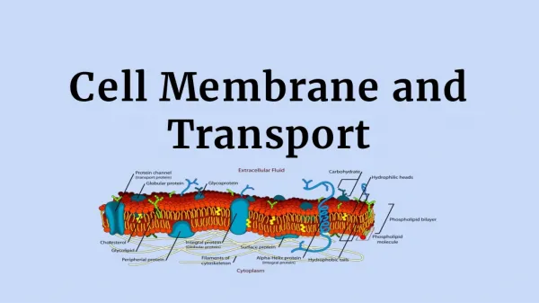
Cell Membrane and Transport. Structure and Function. Plasma Membrane *. Plasma Membrane is made of a bilayer of Phospholipids. A phospholipid has a hydrophilic head and hydrophobic tails, it is amphipathic .

E N D
Cell Membrane and Transport Structure and Function
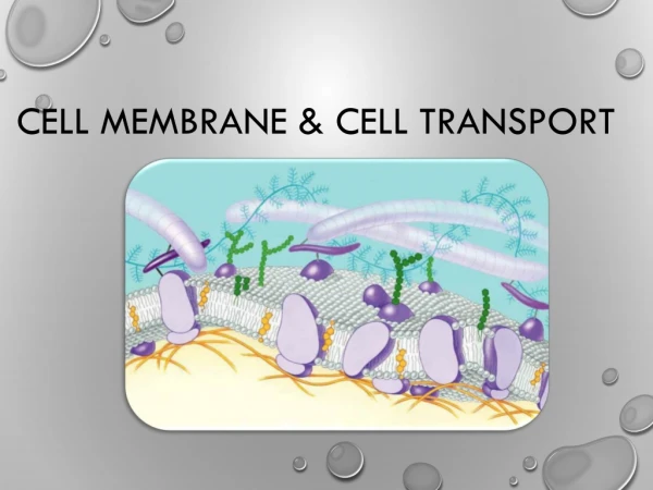
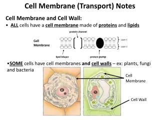
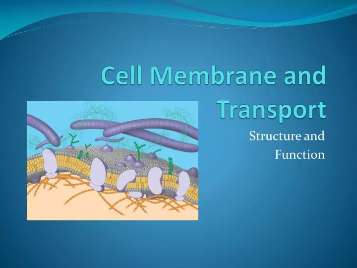
Plasma Membrane * • Plasma Membrane is made of a bilayer of Phospholipids. • A phospholipid has a hydrophilic head and hydrophobic tails, it is amphipathic. • This creates a special dynamic that allows the membrane to maintain an internal environment different from the external and therefore maintain internal homeostasis
Fluid Mosaic Model * 4 Main Components of the plasma membrane contribute to the model’s name. • Phospholipids form the bilayer. • They are viscous and constantly rearranging themselves laterally and flip-flopping this provides the Fluid part of the model. • The unsaturated and saturated tails prevent the phospholipids from tightly packing, or disassociating and falling apart.
Fluid Mosaic Model * 2. Cholesterol is embedded into the bilayer. • It acts as glue reducing the fluidity of the membrane at moderate temps but at lower temps prevent the membrane from freezing. • The cholesterol is a temperature buffer, many winter plants increase the production of cholesterol in the winter to prevent their tissues from freezing.
Fluid Mosaic Model * 3. Proteins are embedded in the bilayer • Proteins give the model its pattern (mosaic). • Proteins have many functions within the membrane: Transport, enzymes, signals, cell to cell recognition and joining • Proteins can be integral/transmembrane or peripheral on the surface only.
Fluid Mosaic Model * 4. Glycolipids and Glycoproteins • These are short polymers of sugars attached to lipids or proteins within the membrane • Function in cell to cell recognition and signaling, organization of tissues, and function of immune system • These biological markers are in a unique pattern on every cell type, and essential to cell recognition ex. A sperm recognizes the ovum, and cells of the immune system recognize bacteria
Synthesis of membranes • Membranes have distinct inside and outside surfaces • Each protein has a directional orientation due to its function • Vesicles are the source of membrane renewal. As they fuse the inside of the vesicle becomes continuous with the outside of the cell. Remember Vesicles are generated by the ER. • So the proteins that end up on the outside of the cell start on the inside surface of the ER.
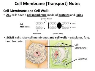
Other specialized structures • Microvilli – extensions of the plasma membrane that increase the absorption area of a kidney or intestinal cell. • Membrane junctions- weave cells together into tissues • Tight junctions- integral proteins of adjacent cells fuse together reducing extracellular space • Desmosomes- create an internal network in which filaments tie cells together to prevent tearing of cellular sheets • Gap junctions- hollow channel in which the plasma membranes of adjacent cells fuse so cytosol can be exchanged important in electrically excitable tissues like the heart and smooth muscle where ion passage helps synchronize activity
Selectively Permeable • The plasma membrane is selectively permeable • Hydrophilic surface and hydrophobic core prevents polar molecules from moving through the membrane. Even water which is very small can only navigate through slowly. • Proteins often provide a path in which molecules can pass into/out of the cell
Passive Transport * • Diffusion is the movement of a substance across a membrane from an area of high concentration to an area of low concentration with out spending energy, Osmosis is the diffusion of water. • Facilitated Diffusion uses Transport Proteins that provide a path for polar molecules and specific ions, each is specific to the substance it helps. • Some are channel proteins that allow substance to move easily through a tunnel down the center, others are carrier proteins that bind with the substance but do not utilize energy in the transport Ex. Aquaporins, are channel proteins specific to water transport, also ion channels, and gated channels Ex. Glucose transporters, are carrier proteins
Diffusion * • Matter is in constant motion, thermal motion. This property drives diffusion. • Molecules move down their concentration gradient until they reach dynamic equilibrium. “High to Low” • Concentration of solutes often drive the diffusion of water, osmosis • Remember the terms solute, solvent, solution?
Osmosis * Tonicity, the ability of a solution to cause a cell to gain or lose water. It depends on the concentration of solutes that can not diffuse across the membrane Isotonic- solute concentrations are same Hypertonic- solute concen. is higher Hypotonic- solute concen. is lower Osmoregulation, the control of water balance A red blood cell placed in: pg 133 isotonic, stays the same, a state of dynamic equilibrim is maintained. hypertonic will shrivel, as water rushed out of the cell. hypotonic will swell/burst open as water rushes into the cell.
Homeostatic balance (not Hbio)* • If a patient has excess fluid in the extracellular spaces a hypertonic solution may be administered to draw the fluid into the blood to then be eliminated by the kidney • If a patient is dehydrated, a hypotonic solution may be given to rehydrate the tissues (sport drinks usually work) • Filtration is a process which forces water and solutes through a capillary cell membrane using a pressure gradient • Blood holds a hydrostatic pressure forcing fluids with vital solutes out to bathe the cells • This occurs in your kidneys, pressure pushes waste solutes out of the blood into kidney where they are collected and eliminated as urine.
Osmosis and Plant cells * Unlike animal cells Plant cells have a cell wall. The cell wall’s main function is support. This is assisted by the central vacuole and internal turgor pressure. A cell that is turgid, rigid, has a high internal turgor pressure, which cancels out the water potential of a hypotonic solution and prevents the cell from bursting. A cell that is flaccid, has lost pressure and may become limp and the plant may wilt. A cell may die if placed in a hypertonic solution because the complete loss of turgor pressure causes plasmolysis, the plasma membrane pulls away from the cell wall and the cytoplasm shrivels.
Active Transport * • Work against a gradient and utilize cellular energy (ATP) • Carrier Proteins are phosphoralized by ATP and change their shape, binding to the molecules they transport across the membrane. Example: Sodium-Potassium Pumpbrings 2 K+ inside and 3 Na+ out (electrogenic pump) • This creates a membrane potential (+ outer surface charge and – internal surface charge) this drives other ion transport, cations in and anions out, this is a chemical force • Together this is called an electrochemical gradient. http://highered.mcgraw-hill.com/sites/0072437316/student_view0/chapter6/animations.html#
Active Transport cont’d • Proton Pump, actively transport H+ out of the cell to generate electrochemical gradient essential in cell respiration and photosynthesis. • Cotransport, uses a proton pump to drive a carrier protein (utilizes H+ gradient instead of ATP) • see handout in packet
Bulk Transport * (see handout in packet) http://www.johnkyrk.com/CellIndex.html Endocytosis • Phagocytosis cellular eating,examoeba,WBC • Pinocytosis cellular drinking, nonspecific intake • Receptor Mediated endocytosis, between the neurons Exocytosis, secretion to export products ex. Neurons release neurotransmitters in bulk
Toxins interupt cellular pathways (not Hbio)* • Toxins from Anthrax, Diphtheria, Tetanus, and bacteria that cause Cholera or Botulism interrupt cell signal pathways by infiltrating the plasma membrane. • Cholera enters an intestinal cell via receptor -endocytosis and rides a “lipid raft” to the ER where it unfolds its deadly proteins causing disease • Botulism toxin causes paralysis by blocking the release via exocytosis, of acetylcholine, the neurotransmitter that links neurons to muscle cells.