Download
1 / 16
160 likes | 253 Vues
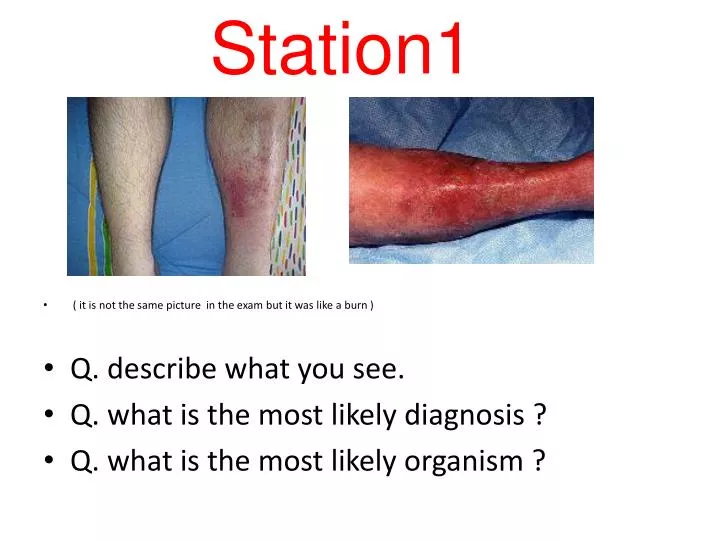
Station1. ( it is not the same picture in the exam but it was like a burn ) Q. describe what you see. Q. what is the most likely diagnosis ? Q. what is the most likely organism ?. describe what you see? a swollen, red area of skin that feels hot and tender, and it may spread rapidly .

E N D
Station1 • ( it is not the same picture in the exam but it was like a burn ) • Q. describe what you see. • Q. what is the most likely diagnosis ? • Q. what is the most likely organism ?
describe what you see? • a swollen, red area of skin that feels hot and tender, and it may spread rapidly. • what is the most likely diagnosis ? • cellulitis • what is the most likely organism ? • Hemolytic streptococci mainly s.pyogenes
Station2 40 yr female , 5 days postpartum!! , she developed swelling & redness in her Rt leg :
a)Dx : b) 2 signs c) 4 risk factors d) 2 medical Rx : e) single best investigation :
2 signs: Dx : DVT. • Redness and warmth over the affected area. • Swelling in the affected leg 4 risk factors? • prior DVT, • obesity, • immobility ( recent major surgery ( abdomial or pelvic, orthopedic surgery, fractures), • OCP post partum period,heriditary , • thrombophilias as factor V leiden ,protein C & S deficiency, malignancies
*2 medical ttt : • TED stockings • graded sequential compression device SCD • Anticoagulation: heparin+warfarin *single best investigation • Venography (gold standard) yes venography is the gold standard but is not the modality of choice and is not the best cuz it is invasive The best would be Duplex ultrasonograghy.Alyaa
Station 3 (picture of lower limbs one is amputated & the other with gangrene) • what is the cause of this operation? • what's the cause? • What are the sign of acute ischemic limb? 4 Signs
Note: amputation can be either above knee(AKA) or below knee(BKA) above knee(AKA) below knee(BKA) • what is the cause of this operation? ulcer &Gangrene in foot
what's the cause? Critical ??Arterial Ischemia (either acute or chronic. We can suspect it’s chronic from the state of the limb, previous ulcers, risk factors for atherosclerosis, hx of intermittent claudication.) • What are the sign of acute ischemic limb? 4 Signs 1) pallor 2) pulselessness 3) paresthesia 4)paralysis –late-
Station 4 • Q. describe? • Q. what is the most likely diagnosis ? • Q. mention 3 findings that you could have by examining such patient.
Q. describe. describe what u see: full description of the ulcer , color, is it wet or dry? The skin near to it.. • Q. what is the most likely diagnosis ? Gangrene • Q. mention 3 findings that you could have by examining such patient. 1)Absent peripheral arterial pulse 2)Cold limbs? 3)Discharge if wet gangrene 4) Absent sensation?(neuropathy)
Station 5 a) Dx b) The affected structure ? c) List 4 complications? d) single surgical treatment?
Dx : varicose veins b) The affected structure : the valves of the long or great sphenous vein ( superficial veins) c) List 4 complications: Most people who have varicose veins will not develop any complications. When complications do develop, it's usually several years after varicose veins first appear. 1)Leg pain 2)Venous ulcers in gaiter’s area. 3)Phlebitis 4)Varicose eczema/ hyperpigmentations 5)lipodermatosclerosis(late):Hardening & tightening of the skin d) single surgical treatment: Vein Stripping (for saphenous vein)
Station 6 Picture of Varicose Vein • Describe? • What symptoms may be associated w/ this condition? • Name which vein may be affected?
Describe: dilated tortuous superficial veins (V.V.) • What symptoms may be associated w/ this condition? • Pain, discomfort, heaviness, itching, skin pigmentation. • Name which vein may be affected?If on the lateral side of the leg: short saphenous if medial aspect: long-great- saphenous Both are superficial veins
Station 7 • : picture 7.15 in browse (describe & give two mechanism) هذي اقرب صورة لقيتها، بس ارجعوا لبراوز أحسن A young woman with primary thrombocythaemia. In addition to occlusion of the hepatic vein, the inferior vena cava was occluded above the right renal vein. An extensive collateral system developed. The photograph shows dilated veins on the anterior abdominal wall connecting the inferior to superior venous systems