Download
1 / 31
380 likes | 927 Vues
Joints of Lower Limb. By Dr.Pardeep Kumar. JOINTS OF LOWER LIMB. Joints of pelvic girdle Sacroiliac joint Bones: auricular surface of sacrum and ilium Capsule: very tight and strengthened by ligaments. Vertebropelvic ligaments

E N D
Joints of Lower Limb By Dr.Pardeep Kumar
JOINTS OF LOWER LIMB Joints of pelvic girdle • Sacroiliac joint • Bones: auricular surface of sacrum and ilium • Capsule: very tight and strengthened by ligaments
Vertebropelvic ligaments • Iliolumbal ligament: runs from transverse process of L5 to the posterosuperior part of iliac crest ★Sacrotuberous ligament: runs from lateral margins of sacrum and coccyx to the inner margin of ischial tuberosity ★Sacrospinous ligament: runs from ischial spine to lateral margins of sacrum and coccyx • These two ligaments convert the sciatic notches the greater and lesser sciatic foramina
Pubic symphysis • Articulation: symphysial surface and interpubic disc (fibrocartilage) • Ligaments: superior pubic ligament and arcuate pubic ligament • Obturator membrane obturator canal
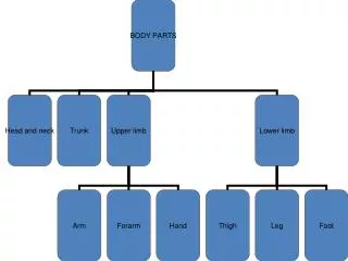
BONY PELVIS Composition: formed by paired hip bones, sacrum, coccyx, and their articulations • In anatomical position, anterior superior iliac spines and pubic tubercles on same vertical plane, while the tip of coccyx and superior border of pubic symphysis on same horizontal plane • Terminal line: formed by promontory of sacrum, arcuate line, pectin of pubis, pubic tubercle, upper border of pubic symphysis
Lesser pelvis • pelvic inlet (terminal line): • Pelvic outlet : formed by tip of coccyx, sacrotuberous ligament, ischial tuberosity, ramus of ischium, inferior ramus of pubic, symphysis • Pelvic cavity • Pubic arch, subpubic angle
Main difference between male and female pelvis Male Female Pelvic inlet Pelvic outet Pelvic cavity Pubic arch 90~1000 70~750
Joints of free lower limb ★ Hip joint • Bones: acetabulum and femoral head • Articular capsule attachments • Above: margins of acetabulum and transverse acetebular ligament • Below: in front to intertrochanteric line; behind, to the neck of femur above 1 cm above the intertrochanteric crest
Acetabulum labrum Ligament of head of femur Transverse acetebular lig. • Accessory structures • Acetabulum labrum; transverse acetebular ligament • Ligaments • Iliofemoral lig. • Ligament of head of femur • Pubofemoral lig. • Ischiofemoral ligament • Zona orbicularis:annular ligament is a ligament on the neck of the femur formed by the circular fibers of the articular capsule of the hip join • Movement: flexion, extention, adduction, abduction, medial and lateral rotation, circumduction
Pubofemoral lig. Iliofemoral lig. Ischiofemoral lig. Zona orbicularis
★Knee joint • Bones: lower end of femur, upper end of tibia and patella • Articular capsule: superapatellar bursa, deep infrapatellar bursa, ala folds
Fibular collateral lig. Patellar lig. Tibial collateral lig. • Accessory structures • ligaments • Patellar lig. • Fibular collateral lig. • Tibial collateral lig.
Oblique popliteal ligament • Anterior cruciate ligment • Posterior cruciateligament
Medial meniscus(C-shaped) • lateral meniscus(O-shaped) • Movements: flexion and extension; flexed knee joint may be passively rotated through 700 lateral Medial
Tibiofibular syndesmosis • Tibiofibular joint • interosseous membrane • Anterior and posterior tibiofibular ligaments
Joint of foot Talocrural joint (ankle joint) • Bones: lower ends of tibia and fibula, trochlea of talus • Articular capsule: thin and lax in front and behind, and supported on each side by strong collateral ligaments
Ligments • Medial lig. • Lateral lig. • Anterior talofibular lig. • Calcaneofibular lig. • Posterior talofibular lig. • Movements: dosiflexion (extension) and plantar flexion (flexion); when the ankle joint is fully plantar flexed, small amounts of abduction, and adduction are possible
Intertarsal joints • Talocalcaneal joint • Talocalcaneonavicular joint • Calcaneocuboid joint • Tarsometatarsal joints • Intermetatarsal joints • Metatarsophalangeal joints • Interphalangeal joints transverse tarsal joint
Arches of foot • Medial longitudinal arch: formed by calcaneus, navicular, three cuneiforms and first to third metatarsal bones, head of talus is the keystone of this arch
Lateral longitudinal arch: formed by calcaneus, cuboid, fourth and fifth metatarsals; cuboid is is the keystone of this arch
Tranverse arch: formed by cuboid, three cuniforms and all metatarsals; the intermediate cuneiform is the keystone of this arch • Function: give foot strength stability and resilience; protect plantar vessels and nerves
Normal arch Flatfoot