Download
1 / 21
210 likes | 217 Vues
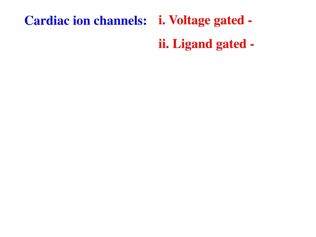
0. I Ca L. I K. mV. -40. -60. Pacemaker potential. i. Voltage gated -. Cardiac ion channels:. ii. Ligand gated -. b) Nodal tissue - RMP & AP. Action potential. I Ca T. 0. mV. -60. 0. mV. Sympathetic stimulation. -60. Vagal stimulation. SYMPATHETIC STIMULATION:.

E N D
0 ICaL IK mV -40 -60 Pacemaker potential i. Voltage gated - Cardiac ion channels: ii. Ligand gated - b) Nodal tissue- RMP & AP Action potential ICaT
0 mV -60 0 mV Sympathetic stimulation -60 Vagal stimulation SYMPATHETIC STIMULATION: PARASYMPATHETIC STIMULATION:
3) CONDUCTIVITY: • SA node: ii. Internodal pathways: iii. AV node: iv. Bundle of HIS: v. Purkinje fibres:
Transtitional cells P - cells i. SA (SINO-ATRIAL) NODE:
ii. INTERNODAL PATHWAYS: • Anterior internodal tract: • Middle internodal tract: • Posterior internodal tract:
SA node Internodal pathways Anterior Middle Posterior
SA node AV node Internodal pathways Anterior Middle Posterior
HIS bundle iii. AV (ATRIO-VENTRICULAR) NODE: N AN NH
iv. BUNDLE OF HIS: • Left bundle branch: • Right bundle branch:
SA node AV node HIS bundle Internodal pathways Rt. branch Lt. branch Anterior Middle Posterior
SA node AV node HIS bundle v. PURKINJE FIBRES: Internodal pathways Rt. branch Lt. branch Anterior Middle Posterior Purkinje fibres
Conduction speeds of different parts (m/sec) Rate of discharge of impulses (impulses/sec) Time taken for impulse to travel different tissues: 0.03sec 0.13sec 0.03sec 0.03sec SA node AV node Bundle branches Purkinje fibers Ventricular endocardial & epicardial surfaces
STEPS IN SPREAD OF ELECTRICAL ACTIVITY: 1. Atrial activation: 2. Septal activation: 3. Activation of: Ventricular myocardium- anteroseptal portion Ventricular myocardium-major portion 4. Late activation of: Left ventricle-posterobasal portion Pulmonary conus Uppermost portion of septum
SA node AV node 1. Atrial activation:
A-V nodal delay- • Duration – • Cause for the delay – • Significance of the delay – • Effect of drugs- • Regulation of A-V conduction: Vagal stimulation Sympathetic stimulation
AV node 2. Septal activation: SA node
AV node 3 (a) Activation of anteroseptal region of ventricular myocardium SA node
AV node 3 (b). Activation of major portion of ventricular myocardium SA node
AV node 4. Late activation of : Posterobasal portion of left ventricle Pulmonary conus & septal uppermost portion SA node
SA AV