Download
1 / 66
660 likes | 803 Vues
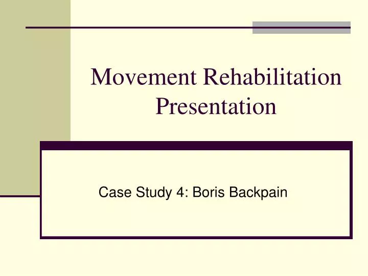
Movement Rehabilitation Presentation. Case Study 4: Boris Backpain. Group Members. Tracy Auld 0275019 Sal Bisignano 0274987 Kylie Knudsen 0275054. Case Study.

E N D
Movement Rehabilitation Presentation Case Study 4: Boris Backpain
Group Members • Tracy Auld 0275019 • Sal Bisignano 0274987 • Kylie Knudsen 0275054
Case Study Boris is a 40 year old male, divorced with 3 children, who live with him every second week and owns a dog. He works as a warehouse manager for an industrial supply company where he administers incoming supplies, stores them and distributes the supplies using truck transportation. Fifty percent of his time he is in the office and the rest out in the warehouse driving a fork lift and directing the loading of materials. Two months ago Boris experienced pain in his lower back, hip and thigh posteriorly after a heavy night on the dance floor with his wife. The next morning he was unable to extend his trunk and had to sit and walk flexed forward. He was taken to the hospital where he was treated with muscle relaxants, anti inflammatory drugs and referred to an orthopedic back specialist. He was referred for an MRI scan which indicated a prolapsed disc but no vertebral abnormality. Musculoskeletal retraining by a physio then an exercise based therapy program was prescribed. This is the second time he has experienced this kind of injury. Boris is 220lbs and 5ft7in. Has been inactive all of his life except in high school. He has worked in physical and manual labor jobs all his life and is naturally strong in the upper body. He is overweight with most of the weight around his mid section. He also likes to eat and cook gourmet meals, drink imported beer, watch football, and fish as a recreation. He is actively involved in transporting his children who are also involved in competitive sport. Boris has completed his treatment for the acute stages of back rehab and is now ready for the development of range of motion and strengthening of the core musculature. He is currently working 3 days a week and experiences fatigue and discomfort at the end of the day. He has also been diagnosed with high blood pressure and high cholesterol, plus he has a family history of heart disease. You have been directed to get Boris back to full function so that he can resume his normal life process.
Background Information • Occupation: warehouse manager • Physical activity level: manual labor work, sedentary on spare time • Physical health: overweight, high blood pressure, high cholesterol • Medical diagnosis: prolapsed intervertebral disc • Physical complaints: pain referral into back, hip and hamstring area • Diet: alcohol consumption high, high saturated fat foods
Rehab Approach • Get Boris back to normal range of motion • Increase his endurance to get through an entire work day • Strengthen core muscles • Approach with prescription of flexibility and strength exercises and functional ability activities
Important Information Before Rehab Begins • Medical history shows that he has high blood pressure and high cholesterol- his doctor must give clearance before exercise is to begin • Medical history also shows that the patient has relapsed in a previous exercise program from a similar injury- motivation is key to success of program • Complete a Par Q (as seen on the next slide) • Allergies? None present. • Medications being taken that pertain to injury and those that do not. Boris is still taking anti inflammatory medications. • Has the patient been released from the rehabilitation with a physiotherapist/occupational therapist. Yes
Communication • The patient must have a say in goal setting. • A therapist must address any fears or personal barriers that may hinder the patient’s recovery. • What are the barriers that the patient is faced with during day to day activities. • How willing is the patient to commit to the prescription in order to recover. • Explain the Borg Scale of Perceived Levels of Pain • Explain the patient’s injury in a way that is comprehensive to the patient.
Borg’s Rate of Perceived Exertion Scale • 1 • 6 No exertion at all • 7 Extremely light • 8 • 9 Very light • 10 • 11 Light • 12 • 13 Somewhat hard • 14 • 15 Hard (heavy) • 16 • 17 Very hard • 18 • 19 Extremely hard • 20 Maximal exertion
Prolapsed Disc The disc that is found in between the vertebrae bones of the back has prolapsed. This means that the gel-like substance, or nucleus pulposus, found in the middle of the disc has been squished out. The area posterior to the disc and bodies of the vertebrae houses the spinal cord. When the disc herniates, the contents of the disc, specifically the nucleus pulposus, are squished into the area where the spinal cord is found. It can also squish into the area where nerves that give rise to sensation in the body exit the spinal canal. If these nerves are constricted, pain in the area where the nerves innervate or weakness may ensue.
Osteology of Effected Area • The vertebral column consists of bone and connective tissue. • Its function is to surround the spinal cord and protect it. • The length of the column is about 71 cm in the average adult male, and 61cm in the average adult female. • It serves as an attachment for the ribs, pelvic girdle, and muscles of the back. • Typically the vertebral column consists of 26 vertebrae. • Vertebrae 1-7 are called the cervical vertebrae. These are the vertebrae of the neck region and attach to the cranium. • Vertebrae 8-19 are called the thoracic vertebrae. These vertebrae provide attachment sites for the ribs. • Vertebrae 20-25 are called the lumbar vertebrae. These vertebrae are found from the end of the ribs until the line where the iliac crests meet. • Inferior to the lumbar vertebrae is the sacrum. The sacrum consists of five fused vertebrae, that are fused in the childhood years. • The final portion of the vertebral column is called the coccyx. • Relative to the front of the body, the cervical and lumbar vertebrae have a convex curve(bulging). • The thoracic and sacral vertebrae have a concave curve(cupping in). • Between the bodies of adjacent vertebrae from the second cervical vertebra to the sacrum are intervertebral discs. • Intervertebral discs consist of an outer fibrous ring composed of fibrocartilage, and an inner soft, highly elastic substance called nucleus pulposus. • These discs form strong joints, which permit various movements of the vertebral column, and absorb compression forces and minimally other related forces of the vertebral column. • Under compression they flatten and broaden. • With age the nucleus pulposus hardens and becomes less elastic.
Myology of Effected Area • Spinalis group: spinalis capitis, spinalis cervicis, spinalis thoracis. Acting together they extend the vertebral column of their respective region. • Transversospinalis group: semi spinalis capitis, semispinalis cervicis, semispinalis thoracis, multifidus, and rotatores. • Semispinalis capitis- acting together they extend the head. Acting individually they rotate the head. • Semispinalis cervicis and semispinalis thoracis- acting together they extend the vertebral column of their respective regions. • Multifidus- Acting together they extend the vertebral column. Acting individually they laterally flex the vertebral column, and rotate the head. • Rotatores- Acting together they extend the vertebral column. Acting individually they rotate the vertebral column. • Segmental group: The segmental group consists of the interspinales. And the intertransversarli. • Interspinales-Acting together , they extend the vertebral column. Acting individually they stabilize the vertebral column during movement. • Intertransversarli- Acting together they extend the vertebral column. Acting individually they laterally flex the vertebral column.
Myology of Effected Area • The Erector spinae is very important in controlling flexion, lateral flexion, and rotation. • The erector spinae is the chief extensor of the back and is the largest muscle mass on the back. • The erectore spinae is composed of three different muscle groups: the iliocostalis group, lingissismus group, and spinalis group. • The erector spinae Originates on: the superior six ribs, inferior six ribs, illiac crest, transverse processes of superior four thoracic vertebrae and articular processes of inferior four cervical vertebrae, transverse processes of 4th and 5th thoracic vertebrae, transverse processes of lumbar vertebrae, ligamentum nuchae and spinous processes of the seventh cervical vertebrae, and spinous processes of superior lumbar and inferior thoracic vertebrae. • The erector spinae inserts on: transverse processes of fourth and sixth cervical vertebrae, superior six ribs, inferior six ribs, transverse processes of second to sixth cervical vertebrae, mastoid process of temporal bone, transverse processes of all thoracic and superior lumbar vertebrae and ninth and tenth ribs, occipital bone, spinous process of axis, and spinous processes of superior thoracic vertebrae.
Neurology of Effected Area • The Spinal cord is the major nerve that innervates the vertebral column. It is roughly cylindrical in shape, but is flattened slightly in its anterior posterior dimension. • In adults the spinal cord runs from the medulla oblongata, to the superior border of the second lumbar vertebra. • The spinal cord is surrounded by extensions of dural sheath called denticulate ligaments. These denticulate ligaments extend all along the length of the spinal cord to support and protect the spinal cord against sudden displacements. • Since the pain the patient is having is in the lumbar region we will focus on the lumbar and sacral nerves that branch out of the spinal cord. • The First group of nerves come from the Lumbar plexus. The lumbar plexus branches off the spinal cord from L1-L4. • The following nerves are the nerves that originate from the lumbar plexus: Iliohypogastric nerve, ilioinguinal nerve, genitofemoral nerve, lateral femoral cutaneous nerve, femoral nerve, obturator nerve. • The next group of nerves that branch off the spinal cord are called the sacral plexus. • The sacral plexus extends from L4-S4. The sacral plexus nerves that branch off the spinal cord between L4-S4. The nerves that originate from the sacral plexus are: superior gluteal nerve, inferior gluteal nerve, the sciatic nerve (branches off into common fibular nerve, and tibial nerve), the posterior cutaneous nerve of thigh, and the pudendal nerve.
Postural Assessment A brief scan of posture should be incorporated prior to range of motion testing. Posterior View • Head: erect or twisted and turned • Shoulders: level or is one slightly higher • Spine: straight or does it appear deviated or scoliotic • Hip: level or is one higher • Feet: pointed straight ahead or outward
Postural Assessment Lateral View • Neck: is it erect or poked forward • Upper back: normally rounded or kyphotic • Trunk: erect or inclined posteriorly • Abdomen: flat or protuding and sagging • Lower back: normally curved or markedly lordotic
Types of Posture • Good • Type A: relaxed, faulty posture • Type B: kyphosis or lordosis • Type C: sway back • Type D: flat back • Type E: round back • Figure 14-3. Types of faulty posture. (From McMorris, R. O.:Pediatr. Clin. North Am. 8:217, 1961.)
Range of Motion Testing of Back Trunk Flexion And Extension Of The Lower Back • The patient stands with feet shoulder width apart. • The patient then flexes the trunk forward to the maximal motion. • A tape measure is used to record trunk flexion and extension. • The therapist finds S2 and measures a point from S2 to 10cm above the S2. Both of these points are marked with a marker. • The patient flexes to his/her maximal point and a measurement is taken between the two lines made previously. • The difference between the two measurements is the lumbar flexion. • Extension can also be measured. The patient has the same two points marked out however, the patient then places his/her hands on the illiac crests and extends to their maximal point. • The difference between these two number is the maximal extension for the lumbar spine. • Normal range is from 0-80º or 10 cm.
Range of Motion Testing of Back Extension of the Thoracolumbar spine • The patient lies prone on the table with a pillow under the abdomen. The hands are positioned at the end of the table. • A strap is placed over the pelvis to stabilize. • The patient extends the elbows to raise the trunk and extend the thoracolumbar spine. • This ROM test is measured with a tape measure. • The tape measure is used to measure the distance between the suprasternal notch and the table at the end of full extension. • Trick movement: Lifting of the pelvis. • Normal range is 0-80º or 10 cm
Range of Motion Testing of Back Trunk Lateral Flexion • The patient stands with the feet shoulder width apart with hands at their side. The patient laterally flexes the trunk to the limit of motion. • A tape measure is used to measure the distance between the third digit and the floor, before the activity and then during activity. • Trick movement trunk flexion, trunk extension, hip and knee flexion. • Normal range is 0-35º
Range of Motion Testing of Back Trunk Rotation • The patient sits on the table with the feet being supported by a stool. • Arms are crossed in front of the chest. • The therapist uses both hands to stabilize the pelvis. • The patient rotates the trunk to the limit of motion. • The measurement used is an estimation by the therapist. • Trick Movement: Trunk flexion, trunk extension, and shoulder abduction. • Normal range is 0-45º
Range of Motion Testing of Hip Hip Flexion PROM Assessment • Patient lies supine on table with the injured leg in a neutral position. The other leg may be flexed or extended. • The therapist stabilizes the pelvis with one hand and places the distal hand on the posterior side where the distal portion of the femur is. • While maintaining stabilization of the pelvis, the therapist applies slight traction to move the femur anteriorly to the limit of hip flexion. • When using the goniometer to measure hip flexion, place the axis over the greater trochanter. The stationary arm should run parallel to the midaxillary line of the trunk. The moveable arm should run parallel to the femur, pointing to the lateral epicondyle. AROM • This assessment is the same as the PROM without assistance • Trick Movement: Posterior tilt of the pelvis and flexion of the lumbar spine. • Normal range of motion is 0-120º
Range of Motion Testing of Hip Hip Extension PROM • The patient lies prone on the table, both hips and legs are in a neutral position with the feet lying over the edge of the table. • The therapist stabilizes the pelvis with the proximal hand while placing the distal hand on the anterior portion of the distal femur. • The therapist then moves the femur posteriorly until full range of motion is achieved. • When using the goniometer the stationary arm lies parallel to the midaxillary line of the trunk. The moveable arm lies parallel to the femur pointing toward the lateral epicondyle. The axis is held over the greater trochanter. AROM • This assessment is the same as PROM without assistance. • Trick movement: The pelvis may tilt anteriorly and extension of the lumbar spine. • Normal range of motion is 0-30º
Range of Motion Testing of Hip Hip Abduction PROM • The patient lies supine on the table with the lower extremities in anatomical position and the pelvis lies level with the lower extremity. • The therapist places the proximal hand on the pelvis to stabilize it, and the distal hand on the medial aspect of the distal femur. • The therapist then moves the femurs away from the body to the limit of abduction. The end feel of hip abduction should be firm. • When using the goniometer the axis is placed over the ASIS of the side being measured.The stationary arm runs along the two ASIS’s. The moving arm is placed anteriorly along the longitudinal axis of femur. • The goniometer measurement will start at 90degrees. Assume 90 degrees is 0 degrees. AROM • This measurement is the same as PROM accept it is without assistance. • Trick Movement: External rotation and flexion of the hip. • Normal range of motion is 0-45º
Range of Motion Testing of Hip Hip Adduction PROM • The patient lies supine on the table with the hip and lower extremities in anatomical position. • The leg that is not being tested is abducted to allow full ROM of the leg being tested. • The therapist places the proximal arm on the pelvis to stabilize it and the distal arm on the posterior aspect of the distal portion of the femur. • The therapist then moves the lower extremity to the limit of hip adduction. • The end feel may be soft or firm. • When using the goniometer the axis is placed over the ASIS of the side being measured. The moving arm is placed anteriorly along the longitudinal axis of femur. • The goniometer measurement will start at 90degrees. Assume 90 degrees is 0 degrees. AROM • This measurement is the same as PROM accept without assistance. • Trick Movement: Internal rotation. • Normal range of motion is 0-30º
Muscle Testing Straight Leg Raising Test • Patient begins by lying in a supine position on the table. • The therapist then passively lifts the patient’s leg by supporting under the calcaneous bone. The other hand is placed over the anterior aspect of the patient’s involved leg in order to maintain a straight leg position. • Lift the leg to approximately 80º • If pain occurs lower the leg and dorsiflex the ankle. • If there is a reaction to dorsiflexion then have the patient locate the pain as precisely as possible. • If there is no pain upon dorsiflexion, • it is an indication of tight hamstrings.
Muscle Testing Well Leg Straight Leg Raise Test • This test is the active version of the straight leg raise test and follows generally the same procedure but with active flexion at the hip. • If back or sciatic pain occurs on the opposite side as being flexed, it is an indication of a prolapsed disc in the lumbar area.
Muscle Testing Hoover Test This test is important because the patient may be less than motivated in performing these tests. The interview prior to the beginning of any program can indicate level of motivation. • The procedure for the well leg straight leg raise test is repeated. The therapist holds both calcaneus bones while the movement is occurring. • If there is no downward pressure being felt on the opposite leg that is being flexed at the hip, the patient is not likely trying.
Muscle Testing Kernig Test This test is used to indicate meningeal irritation, nerve root involvement, and irritation of the dural coverings around the spinal cord. • The patient lays supine and places their hands behind their head. The patient then flexes their cervical spine to produce the action of bringing the chin to the chest. • If there is pain present, it indicates any of the above problems discussed.
Muscle Testing Thomas Test This test is used to indicate whether a patient has hip contractures. Boris has been required to maintain a flexed position for a length of time. • The patient lies supine on the table with the pelvis level and square to the trunk. • The therapist stabilizes the lumbar region by placing their hand under the lumber spine. • The patient flexes the knee and hip to the chest. • The therapist notes the point where the back flattens. This point is important to asses pure hip joint range.
Muscle Testing Thomas Test (cont’d) • The thigh should rest against the abdomen • The patient then flexes the other leg. • Extend the first leg that was flexed onto the table. • If the leg does not rest flat, a contracture may be present.
Expectations of Exercise Prescription • Patient’s goals analysis • Motivation • Weight management • Prevention techniques • Exercise Program • Functional Activities
Patient Goals Analysis What does the patient want to get out of the program? Boris wants to achieve: Full range of motion Increased Endurance Decreased/eliminated pain Reduced body weight Learn how to prevent it
Motivation • Boris has already discontinued one exercise program. • Must find out why the client did not continue with program • Why he quit: • Too busy with work and kids to get to the gym • Too tired after work
Motivation • In order for him to continue with a new program he will have to be convinced that it is worth his time • Benefits of physical activity: • Feel more energetic • Decreased risk of heart disease • Decreased risk of type II diabetes • Weight management • Improved Posture • Sleep better • DECREASE STRESS ON SPINE IN DAILY LIVING!!
Weight Management • Recommendation to nutritionalist • Increase physical activity with exercise program: • Strength Training Program • Walking Program
Prevention Techniques • Understanding Pain Progression • Why is it important? • Understanding the progression of pain or pain patterns associated with the condition will allow for early recognition of reoccurrence and avoidance of activities that cause irritation • Pain associated with a prolapsed disc • Pain tracking depends on the severity of the bugle. Generally the pain starts locally (just around the disc) and migrates down the posterior side of the leg as the condition worsens ( peripheralization). The farther the pain is down the leg, the more severe the bulge is
Prevention Techniques Pain Centralization (condition improving) Pain Peripherilazation (condition worsening) McKenzie Institute International,p 42
Prevention Techniques • Pain During Rehab • During Rehab the pain should migrate out of the leg and into just the back (centralization) • This may make the localized pain in the back feel worse for a while, but ultimately the condition is closer to recovery. • Important: During stretching or physical activity it is important to stop immediately if pain in the leg migrates towards the foot, pain returns in the leg, or localized pain becomes intolerable, as it may be worsening the condition by applying more pressure to the nerve.
Prevention Techniques • Sitting Position • Important to have proper posture when sitting to reduce stress on the back • Proper sitting position is: • Feet flat on the floor • Knees bent at 90 • Back straight and supported Donkin,1986,p 51
Prevention Techniques • Sitting Position continued. . . • Ways to help correct old habits • Back Rest Support (obusform, back roll, towel rolled up) • Strengthen core muscles • Recommendation made that Boris try a back support for his office at work and for his car
Prevention Techniques • Stretching • When sitting for long periods it is important to stretch out your back and your legs • Try to stretch at least every 15 minutes. Donkin 1986,p30 • Every 30 minutes get up to stretch and walk around for a couple of minutes
Prevention Techniques Resting Lying down on your back or lying with your feet elevated takes the compressive forces off of the spine. This will provide some relief for irritated discs. If you have time at noon or on coffee break, take 5 minutes to lie down on a bench or a couch When you get home lie down for 10-15 minutes before driving children or making dinner
Prevention Techniques • Proper Lifting Technique • Be close to the object that is being lifted • Plant feet firmly on the ground in a wide stance • Allow the back to maintain the natural curve of the lumbar spine • Bend the knees to get to the load • Lean back and extend the knees to lift • Lifting should be a smooth movement rather than a jerky one • If performing a rotational movement when under load, turn the feet rather than the back.
Prevention Techniques Proper Lifting Technique McKenzie 2005, p 28
Prevention Techniques • Other Lifting Tips: • Try not to lift heavy loads in the morning when your discs are swollen • Walk around before lifting after sitting • Stretch before and after lifting (standing extensions)
Prevention Techniques • Sneezing/coughing • Sneezing and coughing put a lot of pressure on the discs, especially if they induce forced flexion, and they can be very painful • To reduce pain: • Lean back (put back into extension) • Bend knees
Prevention Techniques • Sleeping soundly • Getting a good sleep is important to everyone’s health. • It can be difficult because of back pain • Ways to minimize discomfort while sleeping: • - Check you mattress. Should be supportive and not sagging • - May want to avoid prone positions • - May want to try a lumbar roll McKenzie 2005, p73