Download
1 / 94
940 likes | 970 Vues
Infrared Spectroscopy. Introduction. Spectroscopy is a technique used to determine the structure of a compound. Most techniques are nondestructive (it destroys little or no sample).

E N D
Introduction • Spectroscopy is a technique used to determine the structure of a compound. • Most techniques are nondestructive (it destroys little or no sample). • Absorption spectroscopy measures the amount of light absorbed by the sample as a function of wavelength.
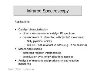
Types of Spectroscopy • Infrared (IR) spectroscopy measures the bond vibration frequencies in a molecule and is used to determine the functional group. • Mass spectrometry (MS) fragments the molecule and measures their mass. MS can give the molecular weight of the compound and functional groups. • Nuclear magnetic resonance (NMR) spectroscopy analyzes the environment of the hydrogens in a compound. This gives useful clues as to the alkyl and other functional groups present. • Ultraviolet (UV) spectroscopy uses electronic transitions to determine bonding patterns.
IR Spectroscopy The entire electromagnetic spectrum is used by chemists: Frequency, nin Hz ~1019 ~1017 ~1015 ~1013 ~1010 ~105 Wavelength, l ~.0001 nm ~0.01 nm 10 nm 1000 nm 0.01 cm 100 m Energy (kcal/mol) > 300 300-30 300-30 ~10-4 ~10-6 g-rays X-rays UV IR Microwave Radio Visible
Infrared Absorbance TV Remote
Infrared Absorbance Sample TV Remote
The IR Region • From right below the visible region to just above the highest microwave and radar frequencies . • Wavelengths are usually 2.5 x 10⁻⁴ to 25 x 10-4 cm. • More common units are wavenumbers, or cm-1, the reciprocal of the wavelength in centimeters. • Wavenumbers are proportional to frequency and energy.
IR Spectroscopy • Used to identify organic compounds • IR spectroscopy provides a 100% identification if the spectrum is matched. • If not, IR at least provides information about the types of bonds present. • Easy to use • liquids analyzed between salt plates • solids in a KBr pellet • small amounts of unknowns via an FTIR microscope • analysis time typically < 10 minutes • Inexpensive • FTIR spectrophotometers are found in most labs.
IR Spectroscopy • IR is used to measure the vibrational frequencies of bonds in the molecule. • Bonds are not rigid. A bond can be viewed as a spring with a weight at each end. • Each bond has a characteristic frequency. • The IR scans a range of frequencies (in the infrared part of the electromagnetic spectrum). Any frequency which matches the characteristic frequency of a bond will be absorbed.
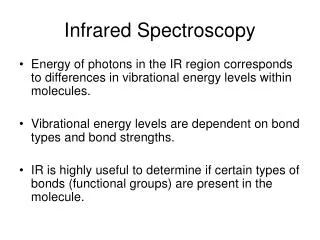
Infrared Spectroscopy • Region of infrared that is most useful lies between 2.5-16 mm (4000-625 cm-1) • IR radiation absorption depends on transitions between Vibrational energy states Stretching: higher energy / higher wave number (cm-1) Bending: lower energy / lower wave number (cm-1)
Vibrations What is a vibration in a molecule? - Any change in shape of the molecule- stretching of bonds, bending of bonds, or internal rotation around single bonds Can a vibration change the dipole moment of a molecule? - Asymmetrical stretching/bending and internal rotation change the dipole moment of a molecule. Asymmetrical stretching/bending are IR active. - Symmetrical stretching/bending does not. Not IR active.
What wavelength of electromagnetic radiation is involved in causing vibrations in molecules? Infrared (IR) electromagnetic radiation causes vibrations in molecules (wavelengths of 2500-15,000 nm or 2.5 – 15 µm) For a vibration at 4111 cm-1 (the stretch in H2), how many vibrations occur in a second? 120 trillion vibration per second!!!! 120 x 1012 vibrations/sec or a vibration every 8 x 10-15 seconds!
A bond must have a dipole or an induced dipole in order to have an absorbance in the IR spectrum. • When the bond stretches, the increasing distance between the atoms increases the dipole moment. Therefore, the greater the dipole, the more intense the absorption. (i.e., The greater the molar extinction coefficient () in Beer’s law, A = bc. )
Molecular Vibrations • If the bond is stretched, a restoring force pulls the two atoms together toward their equilibrium bond length. • If the bond is compressed, the restoring force pushes the two atoms apart. • If the bond is stretched or compressed and then released, the atoms vibrate.
How does the mass influence the vibration? H2 I2 MM =2 g/mole MM =254 g/mole The greater the mass - the lower the wavenumber
Stretching Frequencies • Frequency decreases with increasing atomic mass. • Frequency increases with increasing bond energy.
Effect of an Electric Field on a Polar Bond • A bond with a dipole moment (as in HF, for example) is either stretched or compressed by an electric field, depending on the direction of the field. • Notice that the force on the positive charge is in the direction of the electric field (E) and the force on the negative charge is in the opposite direction.
Vibrational Modes 1. Stretching - the rhythmic movement along a bond axis wit a subsequent increase and decrease in bond length. 2. Bending - a change in bond angle or movement of a group of atoms with respect to the rest of the molecule.
Radiation in the Mid IR region will cause stretching and bending vibrations of the bonds in most covalent molecules. Modes of Vibration: 2. Bending vibrations 1.Stretching Vibrations a. In-plane bending Symmetric stretching Scissoring Rocking b. Out-of-plane bending Asymmetric stretching twisting wagging
Vibrational Modes • A nonlinear molecule with n atoms has 3n - 6 fundamental vibrational modes. • Water has 3(3) - 6 = 3 modes. Two of these are stretching modes, and one is a bending mode (scissoring).
i Number of Vibrational Modes: • - for non-linear molecules, number of types of vibrations: 3N-6 • - for linear molecules, number of types of vibrations: 3N-5 • - why so many peaks in IR spectra • - observed vibration can be less then predicted because • symmetry ( no change in dipole) • energies of vibration are identical • absorption intensity too low • frequency beyond range of instrument Examples: 1) HCl: 3(2)-5 = 1 mode 2) CO2: 3(3)-5 = 4 modes + - - moving in-out of plane See web site for 3D animations of vibrational modes for a variety of molecules http://www.chem.purdue.edu/gchelp/vibs/co2.html
IR Active Vibrations: • - In order for molecule to absorb IR radiation: • vibration at same frequency as in light • but also, must have a change in its net dipole moment • as a result of the vibration Examples: 1) CO2: 3(3)-5 = 4 modes m = 0; IR inactive d- 2d+ d- m > 0; IR active d- 2d+ d- + - - m > 0; IR active d- 2d+ d- degenerate –identical energy single IR peak 2d+ m > 0; IR active d- d-
Mechanical Model of Stretching Vibrations 1. Simple harmonic oscillator •Hooke’s Law (restoring force of a spring is proportional to the displacement) F = -ky Where: F = Force k = Force Constant (stiffness of spring) y = Displacement •Natural oscillation frequency of a mechanical oscillator depends on: a) mass of the object b) force constant of the spring (bond) •The oscillation frequency is independent of the amount of energy imparted to the spring.
Anharmonic oscillators •In reality, bonds act as anharmonic oscillators because as atoms get close, they repel one another, and at some point a stretched bond will break.
•Frequency of absorption of radiation can be predicted with a modified Hooke’s Law. Where: n = wavenumber of the abs. peak (cm-1) c = speed of light (3 x 1010 cm/s) k = force constant m = reduced mass of the atoms Where: Mx = mass of atom x in kg My = mass of atom y in kg •Force constants are expressed in N/m (N = kg•m/s2) -Range from 3 x 10² to 8 x 10² N/m for single bonds - 500 N/m is a good average force constant for single bonds when predicting k. - k = n (500 N/m) for multiple bonds where n is the bond order
Fingerprint Region of the Spectrum • No two molecules will give exactly the same IR spectrum (except enantiomers). • Fingerprint region is between 600–1400 cm-1, and has the most complex vibrations. • The region between 1600–3500 cm-1 has the most common vibrations and we can use it to get information about specific functional groups in the molecule.
Overtones • The vibrations described previously are called fundamental absorptions. (arise from ground state to the first excited state). Usually the spectrum is complicated because of the presence of weaker overtones. • Overtones results from excitation from ground state to the 2nd and 3rd excited states and correspond to integral multiples of the frequency of the fundamental band. Overtones occur at 2 , 3 etc. An absorption in the IR at = 500 cm-1 may have an accomanying peak of lower intensity at = 1000 cm-1 .
Infrared Spectroscopy Infrared Group Analysis: The four primary regions of the IR spectrum Bonds to H Triple bonds Double bonds Single Bonds 4000 cm-1 2700 cm-1 2000 cm-1 1600 cm-1 600 cm-1
Carbon-Carbon Bond Stretching • Stronger bonds absorb at higher frequencies because the bond is difficult to stretch: • C—C 1200 cm-1 • C=C 1660 cm-1 • CC < 2200 cm-1 (weak or absent if internal) • Conjugation lowers the frequency: • isolated C=C 1640-1680 cm-1 • conjugated C=C 1620-1640 cm-1 • aromatic C=C approx. 1600 cm-1
Carbon–Hydrogen Stretching • A greater percent of s character in the hybrid orbitals will make the C—H bond stronger. • An sp3 hybridized carbon has a 25% of S character, an sp2 has around 33% S character, and an sp carbon has 50% S character. • The C—H bond of an sp3 carbon will be slightly weaker than the C—H of an sp2 or an sp carbon.
An IR Spectrum • A plot of % transmittance vs vibrational frequency in wavenumbers (cm-1) λ = wavelength υ = frequency c = speed of light in a vacuum
Wavenumbers • The higher the wavenumber, the shorter the wavelength.
How to Analyze an IR Spectrum • Pay the most attention to the strongest absorptions. • Pay more attention to the peaks to the left of the fingerprint region (>1250 cm-1). • Note the absence of certain peaks. • Be aware of O-H peaks, water is a common contaminant.
IR Spectrum of Alkanes • An alkane will show stretching and bending frequencies for C—H and C—C only. • The C—H stretching is a broad band between 2800–3000 cm-1, a band present in virtually all organic compounds. • In this example, the importance lies in what is not seen, i.e., the lack of bands indicates the presence of no other functional group.
IR Spectrum of Alkenes • The most important absorptions in the 1-hexene are the C═C stretch at 1642 cm-1, and the unsaturated stretch at 3080 cm-1. • Notice that the bands of the alkane are present in the alkene.
O—H and N—H Stretching • Both of these occur around 3300 cm-1, but they look different: • Alcohol O—H is broad with rounded tip. • Secondary amine (R2NH) is broad with one sharp spike. • Primary amine (RNH2) is broad with two sharp spikes. • No signal for a tertiary amine (R3N) because there is no hydrogen.
Characteristic IR Wavenumbers *The peak is broad when H bonding is extensive. Otherwise, the peak can be sharp.
IR Spectrum of Alcohols • The IR spectrum of alcohols will show a broad, intense O—H stretching absorption centered around 3300 cm-1. • The broad shape is due to the diverse nature of the hydrogen bonding interactions of alcohol molecules.
IR Spectrum of Amines • The IR spectrum of amines show a broad N—H stretching absorption centered around 3300 cm-1. • Dipropylamine has only one hydrogen so it will have only one spike in its spectrum.
Carbonyl Stretching • The C═O bond of simple ketones, aldehydes, and carboxylic acids absorb around 1710 cm-1. • Usually the carbonyl is the strongest IR signal. • Carboxylic acids will have O—H also. • Aldehydes have two C—H signals around 2700 and 2800 cm-1.
Variations in C=O Absorption • Conjugation of C=O with C=C lowers the stretching frequency to ~1680 cm-1. • The C=O group of an amide absorbs at an even lower frequency, 1640-1680 cm-1. • The C=O of an ester absorbs at a higher frequency, ~1730-1740 cm-1. • Carbonyl groups in small rings (5 C’s or less) absorb at an even higher frequency. =>
IR Spectrum of Ketones • The spectrum of 2-heptanone shows a strong, sharp absorption at 1718 cm-1 due to the C═O stretch.
IR Spectrum of Aldehydes • Aldehydes have the C═O stretch at around 1710 cm-1. • They also have two different stretch bands for the aldehyde C—H bond at 2720 and 2820 cm-1.