Download
1 / 18
180 likes | 380 Vues
Auditory 2. • Auditory nerve fibers project to the cochlear nucleus, where each fiber splits and goes to each division of the cochlear nucleus (DCN, AVCN, PVCN ). At each step up to the auditory cortex, the tonotopic map is preserved. .

E N D
• Auditory nerve fibers project to the cochlear nucleus, where each fiber splits and goes to each division of the cochlear nucleus (DCN, AVCN, PVCN). • At each step up to the auditory cortex, the tonotopic map is preserved.
The cochlear nucleus is the last place in the auditory system where info from each ear is segregated. • The system has lots of commissures and decussations that allow for communication between the two sides.
• Most other sensory modalities just have a receptors, primary afferent neurons, a primary nucleus, projections to the thalamus and then projections to the cortex. • The auditory system goes from the primary nucleus (cochlear nucleus) to the superior olive, and also to the inferior colliculus before heading to the thalamus. • These two extra centers accommodate the complexity required for sound localization.
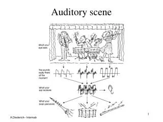
• In addition to sound localization, we have a couple of “de-noising” processes in which we filter our non-essential information. • We direct our auditory attention to particular sounds even when many are present (cocktail party effect).
We also suppress our perception of echoes and pay attention to mostly the first wave front that reaches us (the precedence effect). • For both of these processes, sound localization is important. • Hearing aids often impair these functions.
• The cues for sound localization are the Interaural Time Difference (ITD) with the associated interaural phase difference, and the Interaural Level Difference (ILD). • ITD is due to the pathlength differences to each ear for a sound coming from one side. • These differences are tiny, so require very high time resolution. • ILD is due to the head casting an acoustic shadow on the ear farther from the source.
• The cochlear nucleus outputs are given off by bushy cells. • Bushy cells from both the contralateral and ipsilateral cochlear nucleui give off excitatory fibers to the Medial Superior Olive (MSO) on each side.
Each Lateral Superior Olive (LSO) gets excitatory input from the ipsilateral cochlear nucleus bushy cells, whereas it gets inhibitory input via an MNTB interneuron from the contralateral cochlear nucleus bushy cells.
• Neurons in the auditory nerve and cochlear nucleus fire in phase with the stimulus waveform, called phase locking. • • The MSO is a coincidence detector. • If a sound comes from the right, it takes a little longer to get to the left side.
But, from the left cochlea it has to travel farther to get to the right MSO. • Basically, the time difference it takes for the extra length the AP has to travel across the midline off sets the extra time it takes sound to reach the more distant ear. • So, the signals from each side should arrive at the MSOs on the side the sound comes from at around the same time.
• Depending on the orientation of the head, you’ll get different amounts of time delay (though the neural delay should stay the same). • So depending on the extent of the difference between the two signals, the MSO discharges at different rates.
The MSO is a coincidence detector that responds to how closely the two inputs occur in time. • It converts a time difference into a discharge rate. • By comparing the firing rates of the right and left MSOs, you can get a decent idea of where a sound is coming from.
• Bushy cell (in cochlear nucleus) specializations: Endbulb synapses with the incoming auditory nerve fibers are huge, so when it gets input signals, it sends APs reliably. • They also have a fast membrane time constant, so little temporal summation occurs.
• The LSO gets excitatory input ispilaterally and inhibitory input contralaterally. • It responds to differences in sound intensity between the ears by detecting the balance of excitation/inhibition in each LSO.
• This detection of ITD and ILD well localize the sound to a cone for which these values will be the same. • It doesn’t give you much info on front vs. back or above vs. below, so there must be another cue.
• Spectral cues produced by the external ear resolve this ambiguity. • The pinna reflects the sound and attenuates different frequencies based on where the sound came from along the vertical axis. • The reflections have different pathlengths from the directly incoming sound, and the time delay produces interference that varies with frequency.
So, the frequencies that are attenuated are another cue for where sound comes from. • Thus, it is difficult to localize sound along a vertical axis if you only hear one tone at a time. • You need lots of frequencies to figure out which ones are attenuated.