Download
1 / 43
500 likes | 922 Vues
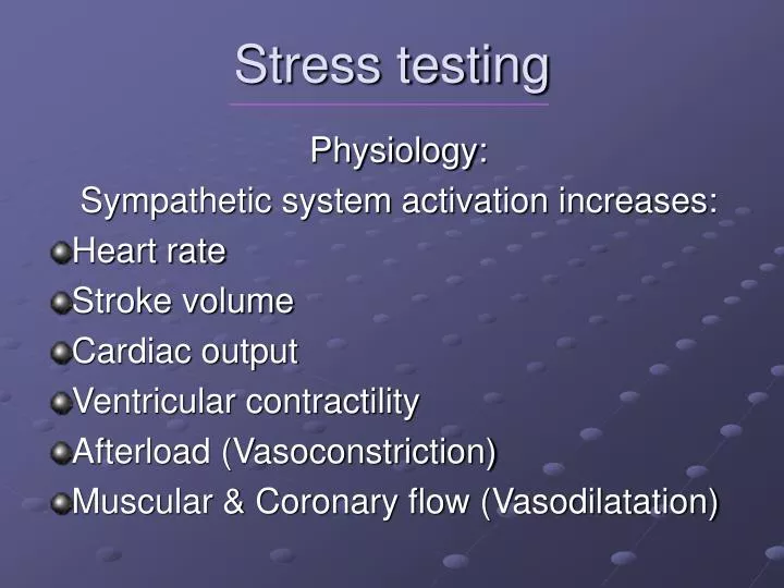
Stress testing. Physiology: Sympathetic system activation increases: Heart rate Stroke volume Cardiac output Ventricular contractility Afterload (Vasoconstriction) Muscular & Coronary flow (Vasodilatation). Demand vs. Supply. Oxygen consumption (VO 2 ). Coronary flow.

E N D
Stress testing Physiology: Sympathetic system activation increases: Heart rate Stroke volume Cardiac output Ventricular contractility Afterload (Vasoconstriction) Muscular & Coronary flow (Vasodilatation)
Demand vs. Supply Oxygen consumption (VO2) Coronary flow . Resting VO2 = 1 Mets = 3,5 ml O2 / min / kg
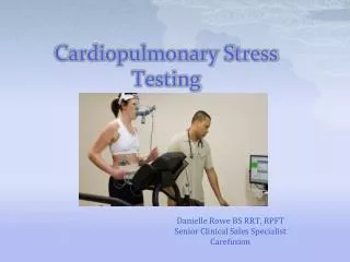
Exercise tests • Master test • Bicycle • Treadmill • ECG - 3 leads (V5), 12 leads • Computerized ST analysis Treadmill stress test
Positive stress test • Anginal pain or dyspnea • ST↓ horizontal >1 mm 0.08” after J point • ST↓ downsloping > 0.5 mm • ST↓ upsloping > 1.5 mm • ST↑ elevation • QRS widening
Exercise test accuracy • Sensitivity =% of pts. w. CAD & ETT(+) ~ 66 % • Specificity = % of normals with ETT(-) ~77 % • False negative: borderline lesions, collaterals • False positive: LVH, MVP, digitalis, LBBB
Indications for ETT I. Diagnostic – probability of CAD • Evaluation of symptoms: chest pain, dyspnea, fatigue • Asymptomatic – Multiple CAD risk factor • Screening • Functional Capacity • Detection of Arrthymia and response to Rx • Hypertensive response
Indications for ETT II. Prognostic: Known CAD – risk stratification • Stable AP, or worsening AP, DOE, FC • Before and after revascularization (PTCA, CABG) • Pre operative risk evaluation
Indications for ETT III. Post Acute Coronary Syndrome • Need for revascularization • Medical treatment adjustment (AP, BP, HR, Arrhythmias) • Guide for cardiac rehabilitation, • Self-confidence • Timing of return to work and its intensity
High risk ETT > 4 % Mortality risk • Low F.C. < 6 min exercise • ST depression at low HR or stress • ST depression > 2 mm • ST elevation or QRS widening • Severe AP or dyspnea • Arrhythmias (VT, PAF) • Systolic BP drop
Contraindications for ETT Risk < 0.01 %, Post MI 0.03% • Unstable Angina • Acute Heart Failure • Arrhythmias • Myo- or Peri-carditis • Severe Aortic Stenosis • Hypertrophic obstructive cardiomyopathy • Severe Hypertension (>220/110 mmHg)
Exercise testing • Fasting, off β-blockers • Symptom limited: AP, dyspnea, dizziness, fatigue, leg pain • Max. heart rate = 220 – age • Target heart rate: 85 % of max. HR If not achieved – non diagnostic test Stop if: ST↓ > 3 mm, ST↑, SBP↓ > 10mmHg, technical problems with ECG monitoring
Nuclear Cardiology Myocardial perfusion Thallium – 201 • Cyclotron product: dose - 2 mCurie • Long half life – 72 hours • 85% - first pass myocardial uptake • Na-K-ATPase pump • Redistribution: 4 or 24 hr.= viability
LAO view of the heart (pathology) A PW S RV LV
Severe exercise – induced ischemia Multiple defects, lung uptake, LV dilatation
Thalium 201 Diagnosis • Infarct: Perfusion defect at stress and rest • Ischemia: Defect at stress that normalizes after 4 or 24 hours. • Sensitivity ~ 90 % • Specificity ~ 80 % • Localization of ischemia / infarct • Extend and severity of CAD • Functional vs. anatomic assessment (angio) • Planar vs. spect (tomographic) imaging
Technetium Sestamibi • Higher dose (30 mCurie), improved image quality • Shorter half life (6 hours) • No redistribution, therefore 2 separate injections for rest and stress • ECG gating for wall motion, EF • First pass imaging
Pharmacologic vs. stress imaging • Indicated for pts. unable to complete full stress test due to low HR, PVD, COPD, CHF, orthopedic disability • Adenosin or dypiridamole drip: vasodilatation of normal vs. narrowed coronaries • Thallium or Tech. sestamibi injection • Perfusion abnormality similar to stress
Contrast left ventricular angiography: Antero – apical aneurysmRAO view Diastole Systole
Technetium 99 labeled RBC m • First pass image or at equilibrium • Multigated acquisition (MUGA) • Regional wall motion at rest and / or stress • Ejection Fraction (%)= X 100 • Assessment of ischemia • Viability: Dobutamine effect EDC - ESC EDC
MUGA – LAO view RV RV LV LV Diastole Systole
Indications for nuclear testing • Diagnostic • CAD assessment – best for intermediate likelihood of CAD • Extent and severity of CAD • Extent of ischemic vs. infarcted areas • Need for revascularization
Indications for nuclear testing II. Prognostic: Risk stratification - MI / Death: 0.5 – 50 % for normal vs. high risk scan • Pre-operative assessment • Post ACS / MI • Change in symptoms / ETT results
High risk nuclear test • Multiple and / or severe perfusion defects • Increased lung uptake • Stress induced LV dilatation
Indications for nuclear testing III. Viability study (hybernating vs. scar tissue) • Thallium late redistribution • MUGA with dobutamine drip • Positron emission tomography (PET) Mismatch between reduced perfusion (ammonia or rubidium) and preserved metabolism (glucose) • Improved function following revascularization