Download
1 / 23
260 likes | 615 Vues
“Clinical Case Challenges in Neuro -Optometry”. Thomas Landgraf, O.D., F.A.A.O. Clinical Associate Professor, UMSL College of Optometry. Neuro -Optometry. Why spend time on it? Conditions are both: Vision threatening Life threatening “True” ocular emergencies. Case #1: ONH Edema?.

E N D
“Clinical Case Challenges inNeuro-Optometry” Thomas Landgraf, O.D., F.A.A.O. Clinical Associate Professor, UMSL College of Optometry
Neuro-Optometry • Why spend time on it? • Conditions are both: • Vision threatening • Life threatening • “True” ocular emergencies
Case #1: ONH Edema? • Always A Tough DDx (Differential Diagnosis) • S: • 52 yo Caucasian male referred to me • Tentative diagnosis of CRVO OS
Case #1: ONH Edema? • Always a Tough DDx • S: • Painless vision loss OS x 2 weeks • Prosthetic OD due to trauma • No significant medical or ocular conditons • Low daily dosage of methadone • Nicotine patch
Case #1: ONH Edema? • Always a Tough DDx • O: • BVA OS: 20/400 • OS pupil round and reactive to light • Normal SLX • Tonometry 17 mm Hg • BP: 280/170 RAS: not done at previous visit
Case #1: ONH Edema? • Always a Tough DDx • O: • DFE OS: • Optic nerve head edema • Accompanied by flame hemes, exudates, cotton wool spots, and macular edema • Normal peripheral retina
Case #1: ONH Edema? • Always a Tough DDx • A: Malignant Hypertension and Resultant Retinopathy OS • P: • Immediate referral to medical center • For lowering of BP • Referral to retinal specialist • Level Of Comfort • Confirmation
Case #1: ONH Edema? • Always a Tough DDx • Follow-up 4 months later • Current meds: minoxidil, norvasc, coumadin • HTN and its complications • Noted improved vision • But some glare, distortion, “wavy lines” in central vision
Case #1: ONH Edema? • Always a Tough DDx • Follow-up 4 months later • BVA OS: 20/20 • BP: 160/85 • DFE OS: exudative macular star, healthy ONH (.2/.2), normal peripheral retina
Case #1: ONH Edema? • Always a Tough DDx • Follow-up 4 months later • Resolving Malignant Hypertensive Retinopathy • Improved Blood Pressure • Educated on compliance
Case #1: ONH Edema? • Always a Tough DDx • Bottom Lines • Primary Care OD’s need to take BP’s • Especially on those with retinal vascular disease • Consider typically bilateral retinal conditions • In monocular patients
Case #1: ONH Edema? • Always a Tough DDx • Timely diagnosis for malignant HTN • Can significantly reduce morbidity and mortality • Like Neuro-Eye Disease: sight and life threatening
Case #2 The BWI Connection • History • 19 yo African-American female • Moderate blur OS • Associated with HA , pain, and pressure OS about 1 month ago • Went to PCP and then referred to eye clinic in DC • Doc did not finding anything wrong…was told it was “sinus-related”
Case #2: BWI • History • Back in Memphis to see family • Mom recommends The Eye Center at SCO • No significant medical or ocular history • Pt is overweight…why mention?
Case #2:BWI • Exam • BCVA: 20/20, 20/25+ • EOM’s: FROM without diplopia • Pupils: grade 1-2 APD OS • Confrontation fields: FTFC OU • Amsler normal OU
Case #2: BWI • Exam • SLX essentially normal OU • IOP: 27, 25 • DFE: • .4/.4 with healthy rim OU • Macula clear OU • Periphery clear OU
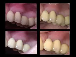
Case #2: BWI • Photos
Case #2: BWI • Ancillary tests ordered • Photos • VF’s: Humphrey 24-2 • Why no optic nerve imaging?
Case #2: BWI • VF OD
Case #2: BWI : “The Clincher” • VF OS
Case #2: BWI • Assessment • 1. Retrobulbar Optic Neuritis OS • Eye pain, APD, central scotoma, decreased VA • 2. Glaucoma suspect OU • Increased IOP • 3. CMA OU
Case #2: BWI • Plan…hmmmm • Patient concerned about cost of visit to neurologist • Wanted to see neurologist when she returned to DC in 2 weeks • Insurance coverage through college • Is this OK?
Case #2: BWI • Plan • 1. Refer to neurologist for further management as deemed appropriate • R/O Multiple Sclerosis • Educated patient on possible etiologies, importance of seeing neurologist, and vision prognosis • 2. Recall in 6 months • 3. No new Rx recommended