Download
1 / 2
20 likes | 118 Vues
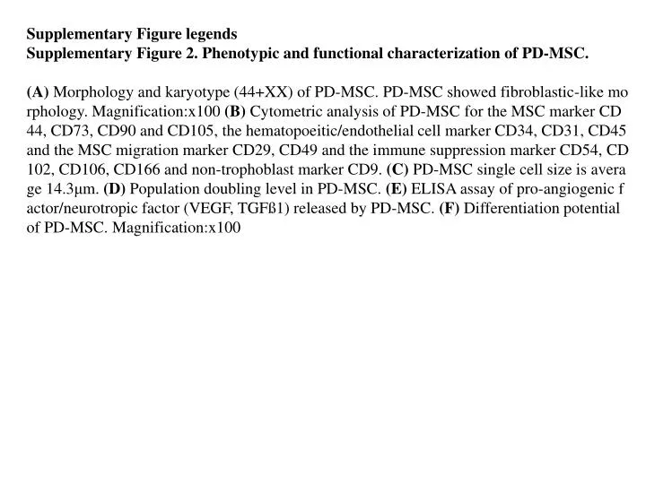
Supplementary Figure legends Supplementary Figure 2. Phenotypic and functional characterization of PD-MSC.

E N D
Supplementary Figure legends Supplementary Figure 2. Phenotypic and functional characterization ofPD-MSC. (A) Morphology and karyotype (44+XX) of PD-MSC. PD-MSC showed fibroblastic-like morphology. Magnification:x100 (B) Cytometric analysis of PD-MSC for the MSC marker CD44, CD73, CD90 and CD105, the hematopoeitic/endothelial cell marker CD34, CD31, CD45 and the MSC migration marker CD29, CD49 and the immune suppression marker CD54, CD102, CD106, CD166 and non-trophoblast marker CD9. (C) PD-MSC single cell size is average 14.3μm. (D) Population doubling level in PD-MSC. (E) ELISA assay of pro-angiogenic factor/neurotropic factor (VEGF, TGFß1) released by PD-MSC. (F) Differentiation potential of PD-MSC. Magnification:x100
B A CD73 CD105 CD44 CD90 CD31 CD29 CD34 CD45 Cell counts CD54 CD166 CD49 CD106 C D SSEA4 HLA-DR CD9 HLA-ABC Fluorescence E F Osteocyte (Von kossa) Chondrocyte (Alcian blue) Adipocyte (Oil red O) CON PD-MSC CON PD-MSC