Download
1 / 76
780 likes | 1.14k Vues
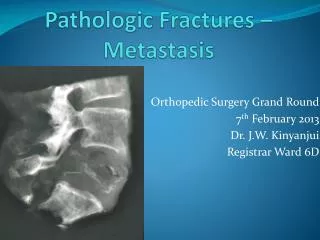
Pathologic Fractures in Children. Joshua Klatt, MD Original Author: Steven Frick, MD; March 2004 1st Revision: Steven Frick, MD; August 2006 2nd Revision: Joshua Klatt, MD; January 2010. Pathologic Fracture = Fracture through abnormal bone. Pathologic Fractures.

E N D
Pathologic Fracturesin Children Joshua Klatt, MD Original Author: Steven Frick, MD; March 2004 1st Revision: Steven Frick, MD; August 2006 2nd Revision: Joshua Klatt, MD; January 2010
Pathologic Fractures • Abnormal bone lacks normal biomechanic and viscoelastic properties • Intrinsic processes • Localized - Bone cyst, neoplasm, etc. • Systemic - OI, osteopenia, osteopetrosis, rickets, etc. • Extrinsic processes • Radiation, biopsy, defects after plate removal, etc.
Osteopetrosis - failed fixation of femoral neck fracture. No osteoclasts = No remodeling.
With every fracture: Ask the question - Is this fracture through NORMAL bone?
Often Need to Do More than Treat the Fracture The orthopaedic surgeon may be the first to have opportunity to make the diagnosis. (malignancy, metabolic disease, etc.)
Often Need to Do More than Treat the Fracture • Differ from fractures in normal bone in that one must take into account… • Etiology • Natural history • Treatment of underlying abnormality • Must treat both fracture and underlying cause!
History • Minor or no trauma? • Less than anticipated for fracture pattern • Any antecedent pain? • Only with activity vs. night pain • Recent illness? • Weight loss? • Fevers?
History • Ask about growth and development • Dietary habits • Kidney disease • May suggest rickets or renal osteodystrophy, etc. • Thyroid disease • Family history • Dysplasias, metabolic disorders, osteoporosis, neuromuscular disorders, etc.
History • Ask about prior malignancies, even in the child! • Families will not always volunteer this information
Physical Exam • Look for soft tissue mass vs. fracture hematoma • Other systems- skin, lymphatics, solid organs • Height - weight percentiles
Lab Tests • CBC with differential • ESR • Calcium (ionized), Phosphorus, Alkaline phosphatase • Bun/Cr
RadiographsBe suspicious! • Osteopenia • Physeal width (rickets) • Soft tissue calcifications • Presence of mass • Any periosteal reaction
Radiographs • Is pathology… • Localized and isolated? • Polyostotic? • Generalized to entire skeletal system? • A generalized condition with skeletal manifestations?
Enneking’s 4 Questions • Where is lesion located? • What is lesion doing to bone? • What is bone doing to lesion? • Are there clues to type of lesion? Enneking, et al. The surgical staging of MSK sarcoma. JBJS 62-A:1027-1030, 1980. Enneking. A System of Staging MSK Neoplasms. CORR 204:9-24, 1986.
Benign vs. MalignantMankin’s Criteria Size Margination Cortex Soft tissue mass Gebhardt, Ready & Mankin. Tumors about the knee in children. Clin Orthop 255:86-110, 1980.
Benign bone lesion Malignant bone lesion Infection Metabolic bone disease Skeletal dysplasia Neuropathic Osteopenia-disuse Overuse Categorize/Make Diagnosis
Union best achieved by correcting biomechanical and biological environment While chemo & radiation slow healing, they provide a beneficial response in presence of rapidly dividing malignant cells Treatment
Simple Bone Cyst(Unicameral Bone Cyst) • Not true neoplasms, etiology unknown • Often loculated and not truly “unicameral” • Most frequently contain serous fluid • Usually metaphyseal • Proximal humeral & femoral lesions account for 94% of all lesions • Most in patients 3-14 years old, average age 9 • Males > females (2:1) Baig & Eady. Unicameral (Simple) Bone Cysts. South Med J. 99(9):966-76, 2006.
SBC Pathologic Fracture • Fallen leaf sign (or fragment) is virtually pathognomonic • Treatment • Fracture heals; cyst persist in 50-90% • Humerus - treat fracture, address lesion after fracture is healed, if felt to be necessary • Displaced proximal femur #s - Open reduction, grafting and rigid fixation, unless very young • Posterior facet #s of the calcaneus - Open reduction, if necessary with grafting and fixation
SBC Treatment • Controversial! • Open Management • Curettage/graft • Bone graft substitutes • Minimally invasive techniques (injections) • Steroid injections • Bone marrow injections • All seem to work with similar frequency (~90%) • But can be recurrence with any of them! • Disrupt hydraulics- puncture, screw, wires, rods, etc.
SBC Injection • 18 ga spinal needle • C-arm • Serous fluid, straw colored • 2nd needle- vent • Depo-Medrol 160 mg • Watch for immediate drainage from large outflow veins • May need multiple injections
LJ, 8 yo with arm pain when throwing, injected once with methylprednisolone (multiple sites), healing at 3 months
SBC - Risk Factors for Recurrence • Only reliable predictor of treatment success is age of the patient • > 10 yrs heal ~ 90% of time • < 10 yrs heal ~ 60% of time • Most cysts tend to heal after skeletal maturity Baig & Eady. Unicameral (Simple) Bone Cysts. South Med J. 99(9):966-76, 2006. Spence et al. Solitary unicameral bone cyst: treatment with freeze-dried crushed cortical-bone allograft. JBJS-A 58:636-41, 1976
Aneurysmal Bone Cyst(ABC) • Expansile • Often wider than physis • Eccentric • Aggressive at margins Cottalorda & Bourelle, Current treatments of primary ABCs. J Pediatr Orthop B 15:155-67, 2006.
Aneurysmal Bone Cyst(ABC) • Symptoms usually present for < 6 months • Lesion may attain considerable size before recognized • Can exist as… • primary bone lesion (70%) • secondary lesion in other osseous conditions (30%) • Pelvic lesions account for 50% of all flat bone lesions (~10% total) • Treatment is difficult due to inaccessibility and integrity of acetabulum Cottalorda et al. Aneurysmal Bone Cysts of the Pelvis in Children. J Pediatr Orthop. 25:471-5, 2005.
ABC Look for fluid-fluid levels on MRI (however, not especially specific) Bur, et al. Fluid-fluid levels in a unicameral bone cyst: CT and MR findings. J Comput Assist Tomogr 17:134-6, 1993. Papagelopoulos, et al. Treatment of aneurysmal bone cysts of the pelvis and sacrum. JBJS-A 83:1674-81, 2001.
5 yo female with 1 year of hip pain and 4 prior steroid injections, progressive coxa vara. Biopsy = ABC
1 month after curettage, bone grafting, valgus/internal fixation, spica immobilization
ABC • Curettage and bone graft • +/- internal fixation • ? Injection of fibrosing agent (Ethibloc, Ethicon, etc.) is controversial • High recurrence Cottalorda & Bourelle, Current treatments of primary ABCs. J Pediatr Orthop B 15:155-67, 2006. Adamsbaum et al. Intralesional Ethibloc injection in primary ABCs. Skeltal Radiol. 32:559-66, 2003. Varshney et al. Is Sclerotherapy Better than Intralesional Excision for treating ABCs. CORR epib 2009.
Nonossifying Fibroma(NOF) • Benign, nonosteoid-producing lesion • Usually found in metaphyses of long bones • Prediliction for lower extremities • Usually asymptomatic • Often incidental radiographic finding • It is speculated that up to a 1/3 of children may have at least a minor NOF/fibrous cortical defect • Almost always regress by early 20’s Betsy et al. Metphyseal fibrous defects. J Am Acad Orthop Surg. 12:89-95, 2004.
Nonossifying Fibroma(NOF) • Most treated non-op! • Let fracture heal, excellent healing potential • Most NOF’s persist after #, but heal by skeletal maturity • If fractures once with minimal trauma, potential risk to fracture again unless bone changes with healing • If necessary, treat with curettage/bone graft Betsy et al. Metphyseal fibrous defects. J Am Acad Orthop Surg. 12:89-95, 2004.
10 yo male - running during soccer. NOF fracture - at 4 weeks underwent allograft DBM / cancellous bone graft. Healed at 9 mos.
NOF - Prophylactic Bone Graft? • Are size parameters predictable? • Arata and Peterson, JBJS 1981 • Review of 23 fractures over 50 years • Suggest tx if greater than 50% diameter, >33 mm length • Easley and Kneisl, JPO 1997 • Review of 22 lesions, many without #s, over 25 years • Only included large lesions (above criteria) • Only 41% had fractures, no refractures • Suggest prophylactic surgery not necessary in many • Criteria for surgery still not well defined Arata et al. Pathological fxs through NOFs. JBJS-A. 63:980-8, 1981. Easley & Kneisl. Pathologic fxs through NOFs: is prophylactic treatment warranted? J Pediatr Orthop 17:808-13, 1997.
Fibrous Dysplasia • Developmental disorder of bone, etiology unclear • May be mutation leading to activation of c-fos oncogene • Can be associated with endocrine disorders (McCune-Albright syndrome) • Can be mono- or polyostotic • Usually affects adolescents and young adults • Many solitary asymp lesions found incidentally • Most do not require intervention • If increased fracture risk, treat with curettage, bone grafting and sometimes internal fixation Parekh et al. Fibrous Dysplasia. J Am Acad Orthop Surg. 12:303-13, 2004.
Fibrous Dysplasia • Surgical treatment for: • Progressive lesions • Large lesions with pain • Pain & deformity suggest microfractures • Failure of conservative treatment • Less successful in: • Younger patients • Larger and proximal femoral lesions • Polyostotic disease, esp McCune-Albright syndrome Enneking & Gearen. Fibrous dysplasia of the femoral neck: Treatment by cortical bone-grafting. JBJS-A 68:1415-22, 1986
Fibrous Dysplasia • Lesions are never eradicated, even with grafting • All grafts are eventually absorbed into dysplastic bone • Cortical grafts at a much slower rate and therefore recommended for weight-bearing bones • Enneking suggested cortical struts alone for femoral neck • Fixation in WB bones seems to improve outcome in children Enneking & Gearen. Fibrous dysplasia of the femoral neck: Treatment by cortical bone-grafting. JBJS-A 68:1415-22, 1986. Stephenson et al. Fibrous dysplasia: An analysis of options for treatment. JBJS-A 69:400-9, 1987.
14 yo female - fell walking across front yard 3 months of left hip pain - Motrin Referred for “path fx through Ewing’s sarcoma” Dx -polyostotic fibrous dysplasia
3 Years Postop Notice resorption of graft and recurrence of cystic changes in femoral neck. Can have secondary ABC develop within fibrous dysplasia.
Fibrous Dysplasia • Consider other sites (polyostotic disease) • Bone scan to help identify other lesions • Although lesions usually revealed on scan, a ‘cold’ bone scan does not rule out lesions • For extensive involvement (McCune-Albright) consider intramedullary fixation/splinting
11 yo male – fem neck path fx, nondisplaced. Fibular allograft (neck) and titanium elastic nails (subtroch and shaft)
13 yrs old – 2 years postop. lesions in Rt. femur and tibia. No pain in hip, in karate.Fibular graft gone. Treat painful tibia? Nail? Pamidronate?
Prophylactic Treatment of Fibrous Lesions (NOF /FD) • Any mechanical pain? • Location and size - relative issues • Supracondylar femur, proximal femur more worrisome • Pharmacologic approach (bisphosphonates) for painful fibrous dysplasia – some reported successes Parekh et al. Fibrous Dysplasia. J Am Acad Orthop Surg. 12:303-13, 2004. DiCaprio & Enneking. Fibrous dysplasia. Pathophysiology, evaluation and treatment. JBJS-A87:1848-64, 2005.
Osteogenesis Imperfecta(OI) • Abnormal type I collagen • COL1A and COL2A defects • Location and type of mutation in collagen molecule determine phenotype (Sillence) • Severe types (II-IV)- multiple fractures prior to skeletal maturity • Also find joint laxity, gray-blue sclera, dentogenesis imperfecta, premature deafness, kyphoscoliosis & basilar invagination • Lower extremity > upper extremity Sillence et al. Genetic heterogeneity in osteogenesis imperfecta. J Med Gen. 16:101-16, 1979. Van Dijk et atl. Classification of Osteogenesis Imperfecta revisited. Eur J Med Genet 53:1-5, 2010.