Download
1 / 25
250 likes | 469 Vues
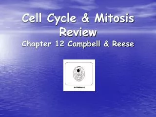
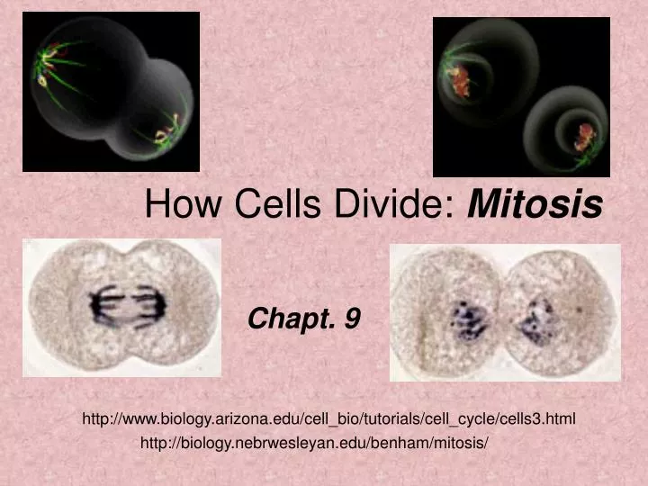
How Cells Divide: Mitosis. Chapt. 9. http://www.biology.arizona.edu/cell_bio/tutorials/cell_cycle/cells3.html. http://biology.nebrwesleyan.edu/benham/mitosis/. Mitosis. Cells must be able to grow & divide New cells must contain the entire set of DNA molecules of the parent cell

E N D
How Cells Divide: Mitosis Chapt. 9 http://www.biology.arizona.edu/cell_bio/tutorials/cell_cycle/cells3.html http://biology.nebrwesleyan.edu/benham/mitosis/
Mitosis Cells must be able to grow & divide New cells must contain the entire set of DNA molecules of the parent cell Cell division involves replication of the DNA molecules stored in chromosomes All the Human chromosomes must be copied and passed to each new cell during mitosis http://www.cellsalive.com/mitosis.htm
Chromosomes:Contain lots of DNA! Chromosome Spread DNA must be highly folded to fit... Some of the DNA from one Chromosome • Text pg 161
DNA folding An average human cell measures ~10µm across…. yet contains ~7 feet of DNA! How to pack it all in??? The DNA double strand is about 2nm across and very long… DNA is highly folded:
ChromatinThe thin, active structure of DNA First level of DNA folding Every 200 nucleotides of DNA wrap around a core of Histone proteins Giving a “beads on a string” look Histones are Basic proteins rich in Amino Acids; Lysine & Arginine -First DNA is wrapped by Histone proteins # 2-4 • Addition of Histone protein 1-Results in a DNA-Histone strand ~30nm wide • Text pg. 161
Chromosome The highly folded DNA structure An inactive form of DNA Chromatin lengths of 50,000-100,000 nucleotides are looped together by nonhistone proteins Chromosomes pack DNA into final structure measuring 5µm long x ~1µm wide • Text pg. 161
DNA Folding An E. coli cell measures about 2µm in length, yet it contains about 1,600 µm (1.6 mm) of DNA --Text pg 156-157 Enough DNA to circle the cell 400 times! A human cell contains enough DNA to encircle it 15,000 times!!! A human cell (35µm diam.) enlarged to 35cm would have 12 miles of DNA Understatement: DNA packaging is important for a cell!
Cell Cycle Cells must be able to grow & divide New cells must contain complete copies of the entire set of chromosomes and all their DNA A Cell’s lifetime of growth & division can be referred to as a Cell Cycle
Cell Cycle Includes not only cell division, but also the intervening time period when cells are not dividing... • Text pg 158
Eukaryotic Cell CycleIncludes: 1. Cell growth 2. Chromosome replication 3. Cell division (2 types) • Mitosis • Meiosis(will discuss later)
Cell Cycle Phases • Interphase: cell growth & DNA replication (steps 1& 2 from previous slide) • Mitosis: nuclear & cell Division
Interphase Composed of G1, S & G2 phases Interphase includes everything except Mitosis
Interphase G1- gap phase between Mitosis & S S phase- DNA replication G2-gap phase between S & Mitosis
Mammalian Cell Cycle G1: Highly variable, Absent in rapidly dividing cells, long in slow-growing cells S: 6-8 hours G2: 3-6 hours M: 1-2 hours
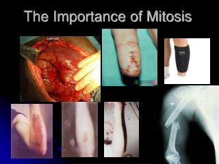
Control of the Cell Cycle For all living eukaryotic organisms it is essential that the different phases of the cell cycle are precisely coordinated. The phases must follow in correct order, and one phase must be completed before the next phase can begin. Errors in this coordination may lead to chromosomal alterations. Chromosomes or parts of chromosomes may be lost, rearranged or distributed unequally between the two daughter cells. This type of chromosome alteration is often seen in cancer cells
2001 Nobel Laureates in Physiology or Medicine Leland Hartwell, Paul Nurse & Timothy Hunt made seminal discoveries concerning the control of the cell cycle. They identified key molecules that regulate the cell cycle in all eukaryotic organisms, including yeasts, plants, animals and human. Defects in cell cycle control may lead to the type of chromosome alterations seen in cancer cells.
From these studies, more than one hundred genes are known to be involved in cell cycle control… known as cell division cycle genes….And that the cell cycle is stopped when/if DNA is damaged. This allows the DNA to repair itself before the cell continues to the next phase of the cycle. One of these key regulators of the cell cycle, CDK (cyclin dependent kinase), controls the cell cycle via the phosphorylation of other proteins. Additional proteins (termed Cyclins) are formed and degraded during each cell cycle. The levels of these proteins vary up and down during the cell cycle. The cyclins bind to the CDK molecules and regulate the activity of CDK activity by selecting the cell proteins to be phosphorylated. These findings led to important insights into how normal and cancer cells develop. • Text pg 159
G1 Arrested Cells • An important control point in cell cycle holds cells in G1 • Cells can remain indefinitely in G1 • Such cells are said to reside in a G0 state, a cell cycle holding point..… • G0 Cells may re-enter the normal cell cycle if given conditions suitable for growth....
The S Phase Each Chromosome replicates to form 2 Chromatids. Replicated chromatids are joined together at their centromeres Replication is semi-conservative. Meaning- each DNA strand serves as a template for a new strand
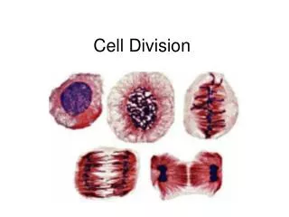
Cell Cycle Phases:M = mitosis Prophase: Metaphase: Anaphase: Telophase:
A. Prophase Chromosome condensation: triggered by changes in histone proteins Nuclear membrane breaks down A microtubular framework (the Spindle apparatus) forms • Text pg 161
B. Metaphase 1. Nuclear membrane breakdown enables spindle microtubules (MTs) to contact chromatids 2. MTs attach to centromere region of each chromatid 3. Chromosomes line up at cell center • Text pg. 163
C. Anaphase • Chromatids are pulled apart by MTs and move toward opposite poles. • Anaphase movement in two parts: • Chromosomes move toward opposite poles • Poles themselves move apart • Anaphase movement uses ATP Text pg. 163
D. Telophase • Chromosomes unfold and disperse (no longer condensed) • Spindle MTs dissassemble • Nuclear membrane Reforms • Gene activity resumes Text pg. 163 Next Step: Cytokinesis
Cytokinesis • Actual cell division stage • In animal cells, a constriction furrow forms on the outside to pinch the new cells apart. • In plant cells, a cell plate forms inside and separates the cells • Text pg. 165