Download
1 / 25
320 likes | 861 Vues
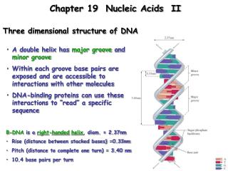
Three-Dimensional Structure of Proteins. Rotation around the a -Carbon in a Polypeptide. A Sterically Nonallowed Conformation. The -Helix and -Pleated Sheet. Conformationally and allowable structures where backbone is optimally hydrogen-bonded (linear H-bonds). -Pleated Sheet:

E N D
The -Helix and -Pleated Sheet Conformationally and allowable structures where backbone is optimally hydrogen-bonded (linear H-bonds) -Pleated Sheet: • anti-parallel or parallel • 2.0 residues/”turn” • 0.34 nm/residue (anti-parallel) or 0.32 nm/residue (parallel) • -Helix: • 3.6 residues/turn • Rise = 0.15 nm/residue • 13-atom hydrogen-bonded loop Linus Pauling and Robert Corey, 1950 Linus Pauling and Robert Corey, 1951
Helices have electric dipoles.
Other Possible Secondary Structures • Helix: • 4.4 residues/turn • 0.12 nm/residue • 16-atom hydrogen-bonded loop • 310 Helix: • 3 residues/turn • 0.20 nm/residue • 10-atom hydrogen-bonded loop
Ramachandran Plot G.N. Ramachandran, 1963
Fibrous ProteinsProteins with an elongated or filamentous form, often dominated by a single type of secondary structure over a large distance. Most fibrous proteins are associated with connective tissue and help provide mechanical strength to the tissue.
Structure of Keratin and Keratin-Type Intermediate Filaments Keratin is a principal component of hair, horn, nails and feathers. Adjacent polypeptide chains also crosslinked by disulfide bonds. Disulfide bond patterns between are what determine whether human hair is straight or curly.
Coiled-Coil -Helical Dimer of a Keratin Amphipathic a helices: Residues a, d, a’ and d’ hydrophobic, other residues more hydrophilic
Structure of Silk Fibroin Silk made by silkworms and spiders. Composed of microcrystalline array of antiparallel pleated sheets where each strand has alternating Gly and Ala or Ser residues.
Structure of Collagen Fibers Collagen is the most abundant vertebrate protein and the major stress-bearing component of connective tissue (bone, teeth, cartilage, tendon) and fibrous matrix of skin and blood vessels. • 3 intertwined left-handed helices • 3.3 residues/turn • Repeating Gly-X-Y (X often Pro, Y often Pro or hydroxyPro)
The Collagen Triple Helix (Tropocollagen) Tropocollagen with Gly Ala substitution (yellow) Interactions between strands G.N. Ramachandran, 1955
Post-Translational Modifications in Collagen Collagen contains unusual oxidized and crosslinked lysine residues. Lysyl oxidase is the enzyme that oxidizes lysine residues to the aldehyde allysine, which then forms the crosslinks. Hydroxyproline is also found in collagen. (Some lysine residues also hydroxylated.) The enzyme required for hydroxylation of proline residues is prolyl hydroxylase, a vitamin C-dependent enzyme. Scurvy is caused by reduced hydroxyproline in collagen as a result of vitamin C deficiency.