Download
1 / 119
1.25k likes | 1.62k Vues
Central Nervous System (CNS). CNS consists of the brain and spinal cord Cephalization Evolutionary development of the rostral (anterior) portion of the CNS Increased number of neurons in the head Highest level is reached in the human brain. Embryonic Development.

E N D
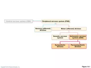
Central Nervous System (CNS) • CNS consists of the brain and spinal cord • Cephalization • Evolutionary development of the rostral (anterior) portion of the CNS • Increased number of neurons in the head • Highest level is reached in the human brain
Embryonic Development • Neural groove fuses dorsally to form the neural tube • Neural tube gives rise to the brain and spinal cord
Regions and Organization of the CNS • Adult brain regions • Cerebral hemispheres • Diencephalon • Brain stem (midbrain, pons, and medulla) • Cerebellum
Cerebral hemisphere Diencephalon Cerebellum Brain stem • Midbrain • Pons • Medullaoblongata (d) Birth Figure 12.3d
Regions and Organization of the CNS • Spinal cord • Central cavity surrounded by a gray matter core • External white matter composed of myelinated fiber tracts
Regions and Organization of the CNS • Brain • Similar pattern with additional areas of gray matter • Nuclei in cerebellum and cerebrum • Cortex of cerebellum and cerebrum
Cortex of gray matter Central cavity Migratory pattern of neurons Inner gray matter Outer white matter Cerebrum Cerebellum Gray matter Region of cerebellum Central cavity Inner gray matter Outer white matter Gray matter Brain stem Central cavity Outer white matter Inner gray matter Spinal cord Figure 12.4
Ventricles of the Brain • Connected to one another and to the central canal of the spinal cord • Lined by ependymal cells
Ventricles of the Brain • Contain cerebrospinal fluid • Two C-shaped lateral ventricles in the cerebral hemispheres • Third ventricle in the diencephalon • Fourth ventricle in the hindbrain, dorsal to the pons, develops from the lumen of the neural tube
Lateral ventricle Septum pellucidum Anterior horn Posterior horn Inferior horn Interventricular foramen Lateral aperture Median aperture Third ventricle Inferior horn Lateral aperture Cerebral aqueduct Fourth ventricle Central canal (a) Anterior view (b) Left lateral view Figure 12.5
Cerebral Hemispheres • Surface markings • Ridges (gyri), shallow grooves (sulci), and deep grooves (fissures) • Five lobes • Frontal • Parietal • Temporal • Occipital • Insula
Cerebral Hemispheres • Surface markings • Central sulcus • Separates the precentral gyrus of the frontal lobe and the postcentral gyrus of the parietal lobe • Longitudinal fissure • Separates the two hemispheres • Transverse cerebral fissure • Separates the cerebrum and the cerebellum
Precentral gyrus Central sulcus Postcentral gyrus Frontal lobe Parietal lobe Parieto-occipital sulcus (on medial surface of hemisphere) Lateral sulcus Occipital lobe Temporal lobe Transverse cerebral fissure Cerebellum Pons Medulla oblongata Fissure Spinal cord (a deep sulcus) Gyrus Cortex (gray matter) Sulcus White matter (a) Figure 12.6a
Central sulcus Frontal lobe Gyri of insula Temporal lobe (pulled down) (b) Figure 12.6b
Anterior Longitudinal fissure Frontal lobe Cerebral veins and arteries covered by arachnoid mater Parietal lobe Right cerebral hemisphere Left cerebral hemisphere Occipital lobe Posterior (c) Figure 12.6c
Left cerebral hemisphere Transverse cerebral fissure Brain stem Cerebellum (d) Figure 12.6d
Cerebral Cortex • Thin (2–4 mm) superficial layer of gray matter • 40% of the mass of the brain • Site of conscious mind: awareness, sensory perception, voluntary motor initiation, communication, memory storage, understanding • Each hemisphere connects to contralateral side of the body • There is lateralization of cortical function in the hemispheres
Functional Areas of the Cerebral Cortex • The three types of functional areas are: • Motor areas—control voluntary movement • Sensory areas—conscious awareness of sensation • Association areas—integrate diverse information • Conscious behavior involves the entire cortex
Motor Areas • Primary (somatic) motor cortex • Premotor cortex • Broca’s area • Frontal eye field
Motor areas Sensory areas and related association areas Central sulcus Primary motor cortex Primary somatosensory cortex Premotor cortex Somatic sensation Frontal eye field Somatosensory association cortex Broca’s area (outlined by dashes) Gustatory cortex (in insula) Taste Prefrontal cortex Wernicke’s area (outlined by dashes) Working memory for spatial tasks Executive area for task management Primary visual cortex Working memory for object-recall tasks Vision Visual association area Solving complex, multitask problems Auditory association area Hearing Primary auditory cortex (a) Lateral view, left cerebral hemisphere Motor association cortex Primary sensory cortex Primary motor cortex Sensory association cortex Multimodal association cortex Figure 12.8a
Primary Motor Cortex Allows conscious control of precise, skilled, voluntary movements
Posterior Motor Anterior Motor map in precentral gyrus Toes Jaw Primary motor cortex (precentral gyrus) Tongue Swallowing Figure 12.9
Broca’s Area • Anterior to the inferior region of the premotor area • Present in one hemisphere (usually the left) • A motor speech area that directs muscles of the tongue • Is active as one prepares to speak
Frontal Eye Field • Anterior to the premotor cortex and superior to Broca’s area • Controls voluntary eye movements
Motor areas Sensory areas and related association areas Central sulcus Primary motor cortex Primary somatosensory cortex Premotor cortex Somatic sensation Frontal eye field Somatosensory association cortex Broca’s area (outlined by dashes) Gustatory cortex (in insula) Taste Prefrontal cortex Wernicke’s area (outlined by dashes) Working memory for spatial tasks Executive area for task management Primary visual cortex Working memory for object-recall tasks Vision Visual association area Solving complex, multitask problems Auditory association area Hearing Primary auditory cortex (a) Lateral view, left cerebral hemisphere Motor association cortex Primary sensory cortex Primary motor cortex Sensory association cortex Multimodal association cortex Figure 12.8a
Primary Somatosensory Cortex • In the postcentral gyri • Receives sensory information from the skin, skeletal muscles, and joints • Capable of spatial discrimination: identification of body region being stimulated
Posterior Sensory Anterior Sensory map in postcentral gyrus Genitals Primary somato- sensory cortex (postcentral gyrus) Intra- abdominal Figure 12.9
Auditory Areas • Primary auditory cortex • Superior margin of the temporal lobes • Interprets information from inner ear as pitch, loudness, and location • Auditory association area • Located posterior to the primary auditory cortex • Stores memories of sounds and permits perception of sounds
Motor areas Sensory areas and related association areas Central sulcus Primary motor cortex Primary somatosensory cortex Premotor cortex Somatic sensation Frontal eye field Somatosensory association cortex Broca’s area (outlined by dashes) Gustatory cortex (in insula) Taste Prefrontal cortex Wernicke’s area (outlined by dashes) Working memory for spatial tasks Executive area for task management Primary visual cortex Working memory for object-recall tasks Vision Visual association area Solving complex, multitask problems Auditory association area Hearing Primary auditory cortex (a) Lateral view, left cerebral hemisphere Motor association cortex Primary sensory cortex Primary motor cortex Sensory association cortex Multimodal association cortex Figure 12.8a
Cingulate gyrus Primary motor cortex Premotor cortex Central sulcus Corpus callosum Primary somatosensory cortex Frontal eye field Parietal lobe Somatosensory association cortex Prefrontal cortex Parieto-occipital sulcus Occipital lobe Processes emotions related to personal and social interactions Visual association area Orbitofrontal cortex Olfactory bulb Primary visual cortex Olfactory tract Fornix Uncus Calcarine sulcus Temporal lobe Primary olfactory cortex Parahippocampal gyrus (b) Parasagittal view, right hemisphere Motor association cortex Primary sensory cortex Primary motor cortex Sensory association cortex Multimodal association cortex Figure 12.8b
Lateralization of Cortical Function • Lateralization • Division of labor between hemispheres • Cerebral dominance • Designates the hemisphere dominant for language (left hemisphere in 90% of people)
Lateralization of Cortical Function • Left hemisphere • Controls language, math, and logic • Right hemisphere • Insight, visual-spatial skills, intuition, and artistic skills • Left and right hemispheres communicate via fiber tracts in the cerebral white matter
Commissural fibers (corpus callosum) Longitudinal fissure Superior Lateral ventricle Association fibers Basal nuclei • Caudate Corona radiata • Putamen • Globuspallidus Fornix Internal capsule Thalamus Gray matter Third ventricle White matter Projection fibers Pons Decussation of pyramids Medulla oblongata (a) Figure 12.10a
Anterior Cerebral cortex Cerebral white matter Corpus callosum Anterior horn of lateral ventricle Caudate nucleus Putamen Lentiform nucleus Globus pallidus Thalamus Tail of caudate nucleus Third ventricle Inferior horn of lateral ventricle (b) Posterior Figure 12.11b (1 of 2)
Cerebral cortex Cerebral white matter Corpus callosum Anterior horn of lateral ventricle Caudate nucleus Lentiform nucleus Thalamus Third ventricle Inferior horn of lateral ventricle (b) Figure 12.11b (2 of 2)
Diencephalon • Three paired structures • Thalamus • Hypothalamus • Epithalamus • Encloses the third ventricle
Cerebral hemisphere Septum pellucidum Corpus callosum Interthalamic adhesion (intermediate mass of thalamus) Fornix Choroid plexus Thalamus (encloses third ventricle) Interven- tricular foramen Posterior commissure Pineal gland (part of epithalamus) Anterior commissure Corpora quadrigemina Mid- brain Cerebral aqueduct Hypothalamus Optic chiasma Arbor vitae (of cerebellum) Pituitary gland Fourth ventricle Mammillary body Choroid plexus Pons Cerebellum Medulla oblongata Spinal cord Figure 12.12
Thalamus • 80% of diencephalon
Thalamic Function • Gateway to the cerebral cortex • Sorts, edits, and relays information • Afferent impulses from all senses and all parts of the body • Impulses from the hypothalamus for regulation of emotion and visceral function • Impulses from the cerebellum and basal nuclei to help direct the motor cortices • Mediates sensation, motor activities, cortical arousal, learning, and memory
Hypothalamus • Forms the inferolateral walls of the third ventricle • Contains many nuclei • Example: mammillary bodies • Paired anterior nuclei • Olfactory relay stations • Infundibulum—stalk that connects to the pituitary gland
Hypothalamic Function • Autonomic control center for many visceral functions (e.g., blood pressure, rate and force of heartbeat, digestive tract motility) • Center for emotional response: Involved in perception of pleasure, fear, and rage and in biological rhythms and drives
Hypothalamic Function • Regulates body temperature, food intake, water balance, and thirst • Regulates sleep and the sleep cycle • Controls release of hormones by the anterior pituitary • Produces posterior pituitary hormones
Epithalamus • Most dorsal portion of the diencephalon; forms roof of the third ventricle • Pineal gland—extends from the posterior border and secretes melatonin • Melatonin—helps regulate sleep-wake cycles
Cerebral hemisphere Septum pellucidum Corpus callosum Interthalamic adhesion (intermediate mass of thalamus) Fornix Choroid plexus Thalamus (encloses third ventricle) Interven- tricular foramen Posterior commissure Pineal gland (part of epithalamus) Anterior commissure Corpora quadrigemina Mid- brain Cerebral aqueduct Hypothalamus Optic chiasma Arbor vitae (of cerebellum) Pituitary gland Fourth ventricle Mammillary body Choroid plexus Pons Cerebellum Medulla oblongata Spinal cord Figure 12.12
Brain Stem • Three regions • Midbrain • Pons • Medulla oblongata
Brain Stem • Similar structure to spinal cord but contains embedded nuclei • Controls automatic behaviors necessary for survival • Contains fiber tracts connecting higher and lower neural centers • Associated with 10 of the 12 pairs of cranial nerves
Frontal lobe Olfactory bulb (synapse point of cranial nerve I) Optic chiasma Optic nerve (II) Optic tract Mammillary body Midbrain Pons Temporal lobe Medulla oblongata Cerebellum Spinal cord Figure 12.14
View (a) Optic chiasma Optic nerve (II) Diencephalon Crus cerebri of cerebral peduncles (midbrain) • Thalamus • Hypothalamus Thalamus Diencephalon Mammillary body Hypothalamus Midbrain Oculomotor nerve (III) Pons Brainstem Trochlear nerve (IV) Medulla oblongata Trigeminal nerve (V) Pons Middle cerebellar peduncle Facial nerve (VII) Abducens nerve (VI) Vestibulocochlear nerve (VIII) Glossopharyngeal nerve (IX) Hypoglossal nerve (XII) Pyramid Vagus nerve (X) Ventral root of first cervical nerve Accessory nerve (XI) Decussation of pyramids Spinal cord (a) Ventral view Figure 12.15a
Crus cerebri of cerebral peduncles (midbrain) Thalamus View (b) Infundibulum Superior colliculus Pituitary gland Inferior colliculus Trochlear nerve (IV) Superior cerebellar peduncle Trigeminal nerve (V) Pons Middle cerebellar peduncle Facial nerve (VII) Inferior cerebellar peduncle Abducens nerve (VI) Vestibulocochlear nerve (VIII) Glossopharyngeal nerve (IX) Olive Hypoglossal nerve (XII) Thalamus Vagus nerve (X) Diencephalon Hypothalamus Midbrain Accessory nerve (XI) Pons Brainstem Medulla oblongata (b) Left lateral view Figure 12.15b
Thalamus View (c) Diencephalon Midbrain • Superiorcolliculus Corpora quadrigemina of tectum • Inferiorcolliculus • Trochlear nerve (IV) Pineal gland • Superior cerebellar peduncle Pons • Middle cerebellar peduncle Medulla oblongata Anterior wall of fourth ventricle • Inferior cerebellar peduncle • Facial nerve (VII) • Vestibulocochlear nerve (VIII) • Glossopharyngeal nerve (IX) Choroid plexus (fourth ventricle) • Vagus nerve (X) • Accessory nerve (XI) Dorsal median sulcus Thalamus Dorsal root of first cervical nerve Diencephalon Hypothalamus Midbrain Pons Brainstem (c) Dorsal view Medulla oblongata Figure 12.15c