Download
1 / 1
10 likes | 78 Vues
Tuberculosis in Badgers: True prevalence, diagnostic methods and epidemiology L.A.L. Corner 1 , D. O’Meara 2 , E. Costello 2 . and E. Gormley 1

E N D
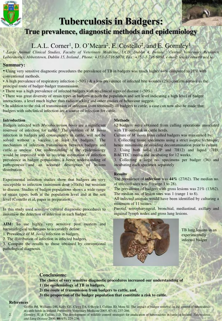
Tuberculosis in Badgers: True prevalence, diagnostic methods and epidemiology L.A.L. Corner1, D. O’Meara2, E. Costello2. and E. Gormley1 1 Large Animal Clinical Studies, Faculty of Veterinary Medicine, UCD, Dublin 4, Ireland. 2Central Veterinary Research Laboratories, Abbotstown, Dublin 15, Ireland , Phone: +353-1-716 6070, Fax: +353-1-716 6091, e-mail: leigh.corner@ucd.ie • Summary: • Using very sensitive diagnostic procedures the prevalence of TB in badgers was much higher 44% compared to 21% with conventional methods. • A high prevalence of respiratory infection (~50%) & a low prevalence of infected bite wounds (2%) confirm aerosol as the principal route of badger-badger transmission. • There was a high prevalence of infected badgers with no clinical signs of disease (~50%). • There was great diversity of strain types in badgers at both the population and sett level indicating a high level of badger interactions, a level much higher than radio-tracking and other studies of behaviour suggest. • In addition to the risk of transmission of infection from terminally-ill badgers to cattle, a case can now also be made that badgers with subclinical infection are a source of infection for cattle. Introduction Badgers infected with Mycobacterium bovis are a significant reservoir of infection for cattle.1 The problem of M. bovis infection in badgers and consequently in cattle, will not be solved without a vaccine, at least for badgers.2 The mechanism of infection transmission between badgers and cattle is unclear. Our understanding of the epidemiology would be improved with an accurate estimation of infection prevalence in badger populations, a better understanding of pathogenesis and an accurate description of lesions distribution. Experimental infection studies show that badgers are very susceptible to infection (minimum dose >10cfu) but resistant to disease. Studies of badger populations shows a wide range of strain types, both at the population level and at the sett level (Costello et al, paper in preparation). In this study used sensitive cultural diagnostic procedures to maximise the detection of infection in each badger. AIM: To use highly very sensitive post mortem and bacteriological techniques to accurately define: 1. Prevalence of M. bovis infection in badgers, 2. The distribution of infection in infected badgers, 3. Compare the results to those obtained by conventional pathological diagnosis. Methods 62 badgers were obtained from culling operations associated with TB outbreak in cattle herds. Culture of M. bovis from culled badgers was maximised by: 1. Collecting tissue specimens using a strict aseptic technique, hence minimising or avoiding decontamination prior to culture 2. Using both solid (LJP and 7H12) and liquid (7H9, BACTEC) media and incubating for 12 weeks. 3. Collecting a large no. specimens per badger (36) and incubating each specimen separately Results The prevalence of infection was 44% (27/62). The median no. of infected sites was 3 (range 1 to 28). The prevalence of badgers with gross lesions was 21% (13/62). The median no. of lesion sites was 1 (range 1 to 8). All infected animals would have been identified by culturing a minimum of 11 tissues: Parotid, retropharyngeal, bronchial, mediastinal, axillary and inguinal lymph nodes and gross lung lesions. TB lung lesions in an experimentally infected badger Conclusions: The choice of very sensitive diagnostic procedures increased our understanding of 1) the epidemiology of TB in badgers, 2) the route of transmission from badgers to cattle, and, 3) the proportion of the badger population that constitute a risk to cattle. • References • Griffin JM, Williams DH, Kelly GE, Clegg TA, O'Boyle I, Collins JD, More SJ. The impact of badger removal on the control of tuberculosis in cattle herds in Ireland. Preventive Veterinary Medicine 2005, 67 (4), 237-266. • Gormley, E. & Collins, J.D. The development of wildlife control strategies for eradication of tuberculosis in cattle in Ireland. Tuberculosis and Lung Disease 2000, 80(4/5), 229-236. • .