Download
1 / 67
700 likes | 1.28k Vues
Vessels and Circulation. Some embryology first. There are at first six pairs of aortic arches In fish these are connected to the gills They undergo a transformation in mammals Birds use the right arch of the fourth pair Mammals use the left arch of the fourth pair. Ventral (anterior) view.

E N D
Some embryology first • There are at first six pairs of aortic arches • In fish these are connected to the gills • They undergo a transformation in mammals • Birds use the right arch of the fourth pair • Mammals use the left arch of the fourth pair
Ventral (anterior) view Transformation : 4th through 7th weeks: some persist, some atrophy Full set of arches develops, but not all present at the same time; (before transformation)
4th arches become: Left side: aortic arch Right side: brachiocephalic trunk Right common carotid a ------------------------------. Right subclavian a. -------------------------- Brachiocephalic trunk-----------------------------------
What the aortic arches become… Right common carotid a ---------------------------. Right subclavian a. --------------------------- Brachiocephalic trunk-------------------------------
3 Major types of blood vessels • Body • RA • RV • Lungs • LA • LV • Boby • Arteries • Capillaries • Veins Arteries carry blood away from the heart -”branch,” “diverge” or “fork” Veins carry blood toward the heart -”join”, “merge,” “converge”
General characteristics of vessels • Three layers (except for the smallest) • Tunica intima - AKA intima • Tunica media – smooth muscle • Tunica externa - AKA adventitia • Lumen is the central blood filled space
Intima is endothelium (simple squamous epithelium) • May have subendothelial layer if 1mm or larger • Tunica media: layers of circular smooth muscles • Lamina (layers) of elastin and collagen internal and external • Thicker in arteries than veins (maintain blood pressure) Smooth muscle contraction: vasoconstriction Smooth muscle relaxation: vasodilation Sympathetic vasomotor nerves of autonomic nervous system regulate
Adventitia (t. externa) – longitudinally running collagen and elastin for strength and recoil
Arteries • Carry blood away from the heart • From big to small, these are the categories: 1. Elastic 2. Muscular 3. Arterioles (then these to capillaries) • Pressure diminishes along the route • Elastic arteries: act as conduits • 2.5-1 cm diameter • Expand with surge of blood from heart • Recoil and continue the propagation of blood • Elastin is thick in media: dampens the surge of blood pressure • Aorta and its branches
Arteries continued 2. Muscular arteries: act as distributing arteries • Middle sized .3mm-1cm • Changes diameter to differentially regulate flow to organs as needed • Internal as well as external elastic lamina • Most of what we see as “arteries” Tunica media larger in proportion to the lumen, thus “muscular”
Arteries continued 3. Arterioles • Smallest: .3mm-10um • Only larger ones have all 3 layers • Regulated 2 ways: • Locally in the tissues • Sympathetic control • Systemic blood pressure (the “BP” we measure) can be regulated through them • Send blood into capillaries Tunica media has only a few layers of smooth muscle cells
Capillaries Heart to arteries to capillaries to veins to heart • Capillaries are smallest • 8-10um • Just big enough for single file erythrocytes • Composed of: single layer of endothelial cells surrounded by basement membrane • Universal function • Oxygen and nutrient delivery to tissues • CO2 and nitrogenous waste (protein break-down product) removal • Some also have tissue specific functions
Capillaries There’s a capillary “bed” in almost all tissues
Capillary permeability • Direct diffusion through endothelial cell membranes • Only O2 and CO2 • Other molecules by various other methods • Blood brain barrier: complete tight junctions • Selective transport of necessary molecules • Lipid soluble agents (like anesthetics) get through, as do O2 and CO2
Veins • Pressure has been lowered so capillaries can tolerate • With lower pressure, walls (of veins) can be thinner • From smallest to large: Capillaries to postcapillary venules to venules to veins • Veins are larger than arteries, plus • Tunica externa is thicker • There is less elastin
Special features of veins • Valves • Prevent backflow • Most abundant in legs (where blood has to travel against gravity) • Muscular contraction • Aids the return of blood to heart in conjunction with valves Mechanical issues… (really good to know)
Exercise helps circulation (because muscles contract and squeeze blood back to the heart)
Vascular anastomoses • Vessels communicating with each other • Veins have more than arteries • Form alternative pathways or collateral channels • Protect organs from being supplied by just one route • Poor anastomoses & therefore vulnerable: central artery of retina, kidneys, spleen, bone diaphyses • Vasa vasorum • Means vessels of the vessels • Blood supply to vessel itself • Smallest vessels don’t need
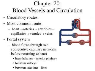
Vascular System (Blood vessels of the body) • Two circulations • Systemic • Pulmonary • Arteries and veins usually run together • Often nerves run with them • Sometimes the systems do not have bilateral symmetry • In head and limbs, most are bilaterally symmetrical
Pulmonary Circulation • Pulmonary trunk branches • Right and left pulmonary arteries • Division into lobar arteries • 3 on right • 2 on left • Smaller and smaller arterioles, into capillaries surrounding alveoli • Gas exchange • Pulmonary system pressure is only 1/6 of systemic blood pressure
Pulmonary Circulation • After gas exchange blood enters venules • Larger and larger into Superior and Inferior Pulmonary veins • Four Pulmonary Veins empty into left atrium
Systemic Circulation • Oxygenated blood to body • Leaves LV through Ascending Aorta • Only branches are the 2 coronary arteries to the heart • Aortic Arch has three arteries branching from it: • Brachiocephalic trunk, has 2 branches: • Right common carotid a. • Right subclavian a. • Left common carotid a. • Left subclavian a. Ligamentum arteriosum connecting to pulmonary a. remember aortic arches…
Descending aorta • Thoracic aorta • at T12 becomes abdominal aorta • Abdominal aorta • ends at L4 branching into: • R & L common iliac arteries
Common carotids branch: • Internal carotids • External carotids • Subclavian: 3 branches • Vertebral arteries • Thyrocerical trunk • Costocervical trunk
Head and neck • Common carotids just lateral to trachea: feel • At larynx divides into internal & external • External carotid: supplies head external to brain and orbit • Feel superficial temporal a. • Middle meningeal: vulnerable (branch of maxillary) • Internal carotid • Supply orbits and most of cerebrum
Internal carotid a. • Enters skull through carotid canal • Gives off: • Ophthalmic artery • Then divides into anterior and middle cerebral arteries (see next slides): together they supply 80% of cerebrum
arteriogram • Middle cerebral arteries run through lateral fissures • Anterior cerebral arteries of each side, through anterior communicating artery, anastomose (an anastomosis is a union)
R and L vertebral arteries* (from subclavians) • Ascend through vertebral foramina of C6-C1 transverse processes • Through foramen magnum into skull • Join to form one Basilar artery* * * * *
Basilar artery: branches • Divides into posterior cerebral arteries • Posterior communicating arteries connect to middle cerebral arteries CIRCLE OF WILLIS Note how it loops around pituitary gland & optic chiasm (now called “cerebral arterial circle”)
Upper limb • Subclavian runs laterally onto 1st rib, under clavicle • Enters axilla as axillary artery • Sends branches • Continues as brachial artery in upper arm • Splits into radial & ulnar arteries • See hand supply Feel brachial & radial pulses
Thorax • Anterior intercostals branch off Internal thoracic*(branch of subclavian) • Posterior intercostals branch off Thoracic aorta Intercostal arteries, veins and nerves run just UNDER the ribs * Small bronchial arteries supply the lung structures
Arteries to the abdomen • Arise from the abdominal aorta • At rest, ½ arterial blood is here! • Three single midline branches supply the digestive tube • Celiac trunk • Superior mesenteric artery • Inferior mesenteric artery 1. 2. 3.
Celiac trunk: divides into 3 right away: left gastric, splenic & common hepatic (see pic; the latter is the only which goes off to the right) • Superior mesenteric supplies most of intestines Definition of mesenteries: double layered sheets of peritoneum that support most organs in the abdominopelvic cavity 1. 2.
(The 1, 2 and 3 are branches of the abdominal aorta) 3. Inferior mesenteric supplies distal half of large intestine 1. 2. 3. Know what these terms mean: phrenic, gastric, hepatic, renal, colic
Arteries to the abdomen • Paired branches off the abdominal aorta supply adrenal glands, kidneys, gonads and abdominal body wall supply diaphragm supply adrenals to kidney 3.
Abdominal aorta branches into Common iliacs at L4; these branch into • Internal iliacs to pelvic organs, perineum, buttocks, medial thighs • External iliacs: to rest of lower limbs
External iliac passes under inguinal ligament becoming Femoral artery • At back of knee femoral becomes popliteal artery, and branches Feel dorslis pedis & posterior tibial