Download
1 / 20
260 likes | 400 Vues
Level 3 Biology. G E N E T I C S. Genetics - Contents. 3 sub topics The role of DNA Gene Expression (the control of) Gene Interactions. Topic 1 - THE ROLE OF DNA. Slide 2 DNA Structure Slide 3 Genes Slide 4 Antiparallel Slide 5 Bases Slide 6 Amino acids

E N D
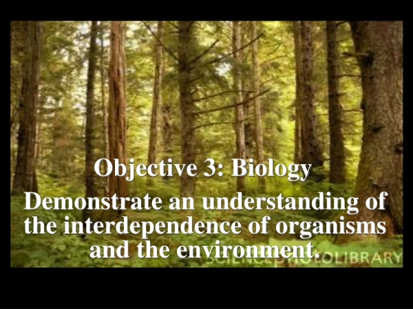
Level 3 Biology GENETICS
Genetics - Contents 3 sub topics • The role of DNA • Gene Expression (the control of) • Gene Interactions
Topic 1 - THE ROLE OF DNA Slide 2 DNA Structure Slide 3 Genes Slide 4 Antiparallel Slide 5 Bases Slide 6 Amino acids Slide 7 Protein Structure Slide 8 Denaturing and types Slide 9 DNA Replication Slide 10 Transcription Slide 11 Translation Slide 12 Prokaryote vs. Eukaryote
What is the difference between prokaryotic chromosomes and eukaryotic chromosomes? • A prokaryotic chromosome consists of a single molecule of DNA in the form of a closed loop. The chromosome is described as circular. • A prokaryotic cell has only one chromosome. • A eukaryotic chromosome is linear, not circular. • Eukaryotic cells have more than one chromosome. • Prokaryotic chromosomes consist only of a naked DNA molecule, • But eukaryotic chromosomes also contain many molecules of proteinsmostlyhistones). The DNA is wound around these proteins. Click image to see full size
DNA STRUCTURE DNA is made of 2 chains of repeating Sugar (deoxyribose) and Phosphate groups. Humans have about 2,900,000, 000 bases (=99cm) in their Genome. See manual pg 53 For this reason it needs to be carefully packaged. It is first coiled, then wrapped around a core of histone proteins. The combined DNA and histone complex is called chromatin, and makes up the structure of a chromatid. Lab manual 55/6
A single gene may have a large number of interruptions in the sequence that codes for a protein. These "introns" are edited out when mRNA is made, but may go on to do a significant function, blowing away the old "one gene, one protein" theory. About 2% of the DNA codes for a protein (the exons). The rest was thought to be "evolutionary junk". Now scientists are finding that much of the rest actually does have a function. 8% of the introns make mRNAchains that have a regulatory purpose. It has now also been found that some exons don’t code for DNA, but mRNA that has a regulatory function. Don’t worry if your confused. More later… Page 68 in Lab manual
Between the 2 chains of DNA are Bases, linked by hydrogenbonds: The 2 strands run in opposite directions (antiparallel). There is a 5' end and a 3' end.
The loss of the 1 oxygen on ribose changes its properties in a very large way. RNA is single stranded. Also in RNA the bases are slightly different… On DNA the bases are Adenine, Thymine, Cytosine, and Guanine. They bond as shown on the right. In RNA Uracil replaces Thymine. Page 69/70 in Lab manual, not 4.
How does DNA form all of that which we are? PROTEIN SYNTHESIS - the process of translating the DNA instructions to make the protein needed. Amino Acids are the building blocks of these proteins. The R side chain is variable, and may be simple (simplest is Glycine where R = H), or very complex. See herefor all of the 20 amino acids used in protein synthesis. 10 of these are "essential amino acids" in our diet, as we cannot make them ourselves. Amino acids link together with a peptide bonds in polymers called Get it? polypeptides.
These polypeptides or polymers or amino acid sequences, call them what you will, form the primary (1°) structure of proteins. The polypeptide folds up in a variety of ways, forming secondary (2°)structures, which are maintained by Hydrogen bonds. Each protein depends on its shape for it's activity (think of enzymes... lock and key... remember?). This distinctive folding pattern is held together mainly by disulfide bridges, formed by cystines interacting. This is the tertiary (3°)structure. Some proteins involve more than 1 polypeptide chain. This is the quaternary (4°) structure.
Proteins, being so dependent on their structure, are prone to damage and subsequent loss of function. This is called denaturing. Denaturing can be caused by: Radiation and heat Heavy Metals Large pH changes Detergents See herefor a narrated animation of an egg denaturing. Page 85/6 in Lab manual (remember to make a half-assed job!) There are 2 structural types of protein:
DNA REPLICATION Have a look at an animation of the process: DNA replication on Youtube What are the main steps and the enzyme catalysing them? Step Enzyme DNA unravelling Helicase Primers added Primase The DNA copied Polymerase Gaps tidied up Ligase Okazaki fragments are made because polymerase only works in the 3’ to 5’ direction. Another way of looking at the same thing: DNA replication 2 Page 77/8 in Lab manual – we don’t need to distinguish between 3b and c. Not 4
Animations • The following animations are provided by the University of Nebraska Institute of Agriculture and Natural Resources. Requires Flash 5 Player. • Transcription/Translation Overview • Transcription Detail • Translation Detail (protein synthesis)
PROTEIN SYNTHESIS • DNA contains the instructions to create every protein used in the body. • The traditional view is that: • 1 section of DNA is copied to make • 1 strand of messenger RNA (mRNA), which moves into the cytoplasm and is used to make • 1 polypeptide chain. • 1 protein is made from 1 or more polypeptide chains. • This is now known to be overly simplistic. Not all DNA is copied. Of the DNA that is copied to mRNA, not all of it is turned into proteins. However, some of the resulting mRNA has a function. • Lets start with the simplest example: Page 87/88/(75) in Lab manual
PROTEIN SYNTHESIS 1. TRANSCRIPTION The process where the DNA strand is copied onto a strand of mRNA. (m=messenger) The DNA strand unravels slightly… The DNA has a Coding strand and a Template (non-coding) strand. The formation of mRNA on the Template is catalysed by RNApolymerase. Triplets on the DNA template code for codons on the mRNA strand. Triplet Codon T A C A U G Page 91 in Lab manual
2. TRANSLATION Transfer RNA (tRNA) molecules move into a ribosome, where they line up with the mRNA. The codons from mRNA line up with anticodons on the tRNA molecule. Amino acid attachment site Anticodon loop mRNA codon table Animationof Protein Synthesis - Translation - (more complex than we need). Page 92-3 in Lab manual
GENE EXPRESSION DIFFERENCES IN PROKARYOTES AND EUKARYOTES Bacteria don’t have a nucleus. This makes protein synthesis a bit easier as mRNA doesn’t have to travel from the nucleus to the cytoplasm as in eukaryotes. Transcription and translation happen at the same time. Also there are very few introns in Prokaryotic DNA. This makes the whole process much more simple. In your average eukaryote the mRNA must be processed in the nucleus to separate the introns and the exons, and the exons reassembled, before translation takes place in the cytoplasm. Check out this for an animation on the differences. Pages 89-90 in Lab manual