Download
1 / 38
400 likes | 433 Vues
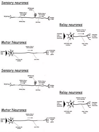
Neurones. Schwann cells form myelin sheath. Types of neurone. There are several different types of neurone:. sensory neurones:. receptor organs. CNS. relay neurones:. motor neurones. sensory neurones. motor neurones:. effectors. CNS. Structure of neurones.

E N D
Schwann cells form myelin sheath Coordination & the Nervous system
Types of neurone There are several different types of neurone: sensory neurones: receptor organs CNS relay neurones: motor neurones sensory neurones motor neurones: effectors CNS
Structure of neurones Cell body – nucleus and large amounts of RER associated with production of proteins and neurotransmitter. Dendrons (dendrites) – carry nerve impulses towards the cell body Axon – single long fibre that carries nerve impulses away from the cell body Schwann cells – surround axon by wrapping around many times, protecting it and providing electrical insulation.
Structure of neurones Myelin sheath – forms covering of axon and made of membranes of the Schwann cells. Rich in a lipid known as myelin. Can be myelinated or unmyelinated. Myelinated neurones transmit nerve impulses faster. Nodes of Ranvier – gaps between adjacent Schwann cells where there is no myelin sheath. Gaps 2-3um and occur every 1-3mm
Transverse section through an axon showing the myelin sheath
The membrane of the neurone contains carrier and channel proteins that are essential to the transmission of nerve impulses. Now answer questions 1-3 on your sheet.
Nerve impulse A nerve impulse is a self propagating wave of electrical disturbance that travels along the surface of the axon membrane. It is not an electrical current, but a temporary reversal of the electrical potential difference across the axon membrane. This reversal is between 2 states called the resting potential and the action potential.
What is a nerve impulse? All cells have a difference in electrical charge across the plasma membrane. This is called the potential difference (p.d.). It is measured in millivolts (mV). plasma membrane axon microelectrodesmeasuring p.d. The normal, resting state of an axon is called the resting potential. In this state the p.d. across the axon is –70mV. The membrane is said to be polarized. A nerve impulse, or action potential, occurs when the p.d.across an axon is temporarily reversed. The p.d. changes to around +35mV. The membrane is said to be depolarized.
Resting potential The establishment of the potential difference (the difference between the inside and the outside of the axon) is due to the following: • Na+ ions are actively transported out of the axon by the Na+-K+ pump. • K+ ions are actively transported into the axon by the Na+-K+ pump.
Resting potential • 3 Na+ move out for every 2 K+ ions that move in. • Results: • more Na+ ions in the tissue fluid surrounding the axon than in the cytoplasm • more K+ ions in the cytoplasm than in the tissue fluid. This creates a chemical gradient.
Resting potential • The Na+ ions should diffuse back into the axon whilst the K+ ions should diffuse out of the axon (through gated channel proteins). • However: • Most K+ channels are open • Most Na+ channels are closed. The axon membrane is 100 times more permeable to K+ ions than Na+ ions.
Resting potential • This increases the potential difference (difference in charge) – the tissue fluid has a positive charge and the inside of the axon has a negative charge due to large proteins etc. (an electrical gradient). • This makes outward movement of K+ ions difficult because they are repelled by the positive charge of the tissue fluid and attracted to the negative charge of the axon, which prevents them from moving out of the axon.
Resting potential 9. An equilibrium is established in which the chemical and electrical gradients are balanced and there is no net movement of ions. At this point, the inside of the axon has a charge of -70mV. This is the resting potential.
The action potential When a stimulus is received by a receptor, its energy causes a temporary reversal of the charge on the axon membrane. As a result, the negative charge of -70mV inside the membrane becomes a positive charge of around +40mV. This is known as the action potential, and in this condition the membrane is said to be depolarised.
The action potential The depolarisation of one section of neurone triggers the next section to become depolarised. This is the wave of depolarisation which we have referred to as a ‘nerve impulse’.
The action potential • At resting potential some K+ voltage-gated channels are open but Na+ channels are closed. • Energy of stimulus causes some Na+ voltage-gated channels in the axon membrane to open. • Na+ ions diffuse into the axon along their electrochemical gradient.
The action potential • As the Na+ diffuse into the axon, more Na+ channels open. The charge inside the axon increases to around +35mV (depolarisation). • The depolarisation of one section of axon membrane stimulates Na+ voltage-gated ion channels to open, allowing the next section of membrane to depolarise.
The all or nothing principle For an action potential to be generated, the stimulus must be greater than the threshold value. 40 0 failed initiations A stimulus will be below the threshold value if insufficient numbers of sodium channels open, preventing full depolarization of the axon. potential difference (mV) thresholdvalue –70 0 1 2 3 4 5 time (ms) Once the threshold value is reached, the action potential generated is always the same size regardless of the strength of the stimulus. It is an ‘all or nothing’ response.
The action potential - repolarisation • The increased voltage causes the voltage-gated K+ channels to open. Na+ voltage-gates close after about 0.5ms. • K+ ions now leave the axon, causing it to repolarise. • Diffusion of K+ ions causes a temporary overshoot of the electrical gradient, (the axon becomes more negative than usual) (hyperpolarisation).
The action potential - repolarisation • The gates on the K+ channels now close and the activities of the Na+ - K+ pumps once again cause Na+ to be pumped out and K+ to be pumped in. • The resting potential of -65mV is re-established and the axon is said to be repolarised.
Passage of the Action Potential Once it has been created, an action potential ‘moves’ rapidly along the axon. The size of the action potential remains the same from one end of the axon to the other. The movement is the reversal of electrical charge at different point along the axon membrane.
Passage of the Action Potential As one region of the axon produces an action potential and becomes depolarised, it acts as a stimulus for the depolarisation of the next region of the axon. Action potentials are regenerated along each small region of the axon membrane. In the meantime, the previous region of the membrane returns to its resting potential, that is it undergoes repolarisation.
What affects the speed of an impulse? An action potential moves faster along myelinated neurones than those without a myelin sheath. Ion channels present at the nodes of Ranvier allow the movement of sodium and potassium ions across the membrane. It is at these points that an action potential can be generated in a myelinated neurone. The action potential moves from node to node.This is referred to as saltatory conduction and is much quicker than the step-by-step conduction that occurs in a non-myelinated neurone.
Myelinated neurones… SALTATORY CONDUCTION
What affects the speed of an impulse? The speed of a nerve impulse is also affected by: • axon diameter – The greater the diameter of the axon the faster the impulse travels. Axons with a small diameter have a larger surface area to volume ratio than axons with a large diameter. This causes a larger number of ions to leak out of the axon, making it more difficult for an action potential to propagate. • temperature – The higher the temperature, the faster the speed of the impulse. Temperature affects the rate of diffusion of ions across the axon.