Download

1 / 97
1k likes | 1.44k Vues
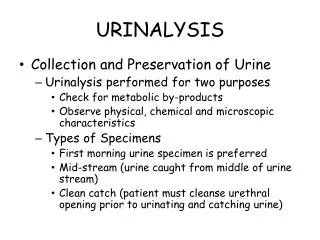
Reference: Laboratory Procedures for Veterinary Technicians 5 th Ed. (Hendrix, Sirois ). Urinalysis. Label samples immediately after collection and perform u/a as soon as possible. Keep reagent strips and tablets in tightly sealed bottles; replace outdated reagents with fresh reagents.

E N D
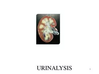
Reference: Laboratory Procedures for Veterinary Technicians 5th Ed. (Hendrix, Sirois) Urinalysis
Label samples immediately after collection and perform u/a as soon as possible. Keep reagent strips and tablets in tightly sealed bottles; replace outdated reagents with fresh reagents. Controls are available to verify strange results. U/a report should include patient information, collection technique, date and time collected, method of preservation (if used) and complete results. Quality Assurance
Analyze within 1 hour to avoid postcollection artifacts and degenerative changes. If immediate analysis not possible, refrigerate for 6-12 hours max. Allow refrigerated sample to come to room temp. prior to evaluation Mix sample well to evenly distribute formed elements. Specimen storage and handling
Samples allowed to stand at room temp. for long periods of time: • Decreased glucose & bilirubin • Increased pH • Crystal formation • Increased turbidity • Breakdown of casts & RBCs • Bacterial proliferation Specimen storage and handling
When sending sample to lab or if sample must be held for >12 hrs, you can add: • 1 drop of 40% formalin in 1 oz. of urine • Toluene sufficient to form a layer on top of sample • A single thymol crystal • One part 5% phenol to 9 parts urine When sending samples to outside lab
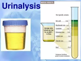
Include all observations made without the aid of microscope or chemical reagents Volume, color, odor, transparency, and specific gravity (spG) of the urine are evaluated Physical properties of urine
May be affected by factors unrelated to disease: • Fluid intake • External losses • Respiratory, intestinal tract • Environmental temperature and humidity • Amount and type of food • Level of physical activity • Size of animal • species Urine volume
Observing a single urination is not reliable for estimating urine output • Ideally determine 24-hour urine volume • Daily urine production varies in different domestic species • Normal daily output = 20-40 mg/kg (dogs and cats) • Terms: • Polyuria, polydipsia, oliguria, anuria Urine volume
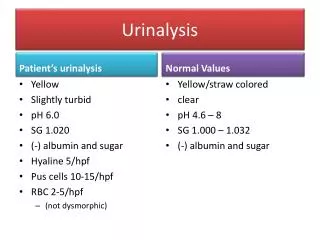
Normal urine = light yellow to amber • Due to presence of urochromes • Magnitude of yellow color varies with degree of urine concentration or dilution • Colorless urine usually has low spG and may be associated with polyuria • Dark yellow to yellow-brown urine generally has high spG and may be associated with oliguria Urine color
Yellow-brown, green, or greenish-yellow, foamy urine contains bile pigments Red or red-brown urine indicates hematuria or hemoglobinuria Brown urine may contain myoglobin Some drugs may alter urine color Evaluate urine for color in a clear plastic or glass container against a white background. Urine color
In most species, freshly voided urine is transparent or clear • Normal equine urine is cloudy; rabbit urine = milky • When observing urine for degree of transparency, place it against a letterprint background. • Transparency is noted as clear, slightly cloudy, cloudy, or turbid (flocculent) Clarity/transparency
Urine may become cloudy while standing because of bacterial multiplication or crystal formation Substances that cause urine to be cloudy include: RBCs, WBCs, epithelial cells, casts, crystals, mucus, fat, and bacteria Other causes of turbidity can include contaminants from the collection container or surface and contamination with feces. Clarity/transparency
Normal urine has a distinctive odor that varies among species Urine of male cats, goats, and pigs has a strong odor An ammonia odor may occur with cystitis caused by bacteria that produce urease(Proteus spp. or Staphylococcus spp.) Samples left standing at room temp. may develop an ammonia odor from bacterial growth Odor
A characteristic sweet or fruity odor to urine indicates ketones and is most commonly found with: • Diabetes mellitus • Ketosis in cows • Pregnancy disease in sheep • postparturent ketosis, eclampsia odor
SpG = weight (density) of a quantity of liquid compared with that of an equal amount of distilled water. Number and molecular weight of dissolved solutes determine SpG of urine. SpG may be determined before or after centrifugation because the particles that settle during centrifugation have little or no effect on SpG. Specific gravity
If urine is turbid, best to centrifuge and then use supernatent to determine SpG. • SpG of normal urine = variable; depends on: • Eating and drinking habits • Environmental temperature • Time of sample collection • Early morning, mid-urination sample tends to be most concentrated. Specific gravity
Interpretation of urine spG yields information on hydration status and the ability of the kidneys to concentrate or dilute urine. • Most commonly determined by refractometer • Reagent strips = least reliable method • Normal ranges: • Dog = 1.001 – 1.060 (1.025) • Cat = 1.001 – 1.080 (1.030) Specific gravity
Increased spG is seen with decreased water intake, increased fluid loss through sources other than urination (e.g. sweating, panting, diarrhea), and increased excretion of urine solutes. Decreased spG is seen in diseases in which the kidneys cannot resorb water and with increased fluid intake, such as polydipsia or excessive fluid administration. Specific gravity
Testing for various chemical constituents of urine is performed with reagent strips impregnated with appropriate chemicals or reagent tablets. Be aware of expiration dates Some reagent strips test for numerous constituents simultaneously; others exist for individual tests Urine is added to reagent strip via pipette or the strips are dipped in the urine sample and color changes are noted at specific intervals. Chemical properties
pH is a measure of hydrogen ion concentration • Above 7.0 = alkaline; below 7.0 = acidic • Normal pH (dog and cat) = 6-7 • If too acidic or too alkaline, specific crystals or uroliths can form • pH of samples left standing open at room temp. tends to increase from loss of CO2 • Delays in reading reaction may lead to color changes and false readings pH
pH of a healthy animal’s urine depends largely on diet. Alkaline urine usually found in animals on plant diets; high-protein cereal diets or diets of animal origin cause acidic urine. Herbivores normally have alkaline urine; carnivores acidic urine; omnivores either acidic or alkaline depending on what was ingested. pH
Usually absent or present only in trace amounts in normal urine obtained by catherization or cystocentesis Voided samples or those obtained by expressing the bladder may contain small amount of protein resulting from secretions that may contaminate urine during its passage along the urinary tract. Protein
Trauma to urinary tract from cystocentesis, catheterization, or bladder expression may cause sufficient bleeding to cause a trace of protein in the urine. Protein levels in urine can be measured by reagent test strips, sulfosalicyclic acid turbidity test, and urine protein/creatine ratio (UPC). protein
Reagent strips (dipsticks) measure protein by progressive color changes on the reaction pad. • Primarily detect albumin • False positive results may occur in alkaline urine depending on diet, urinary tract infection, or urine retention (urethral obstruction) • UPC ratio can help confirm significant amounts of protein in urine • Sample is centrifuged and supernatent is used • Ratio is obtained by dividing protein concentration by creatinine concentration Protein
Very dilute urine can yield false-negative because the concentration may be below the sensitivity of the testing method. • Transient proteinuria may result from a temporary increase in glomerular permeability, allowing excessive protein to enter filtrate • May be found with muscle exertion, emotional stress, or convulsions • Occasionally a small amount of urine protein is found after parturition, during the first few days of life, and during estrus. protein
In most cases, proteinuria indicates disease of urinary tract, especially the kidneys Multiple myeloma, a cancer of plasma cells, may produce large quantities of protein Proteinuria of renal origin may be caused by trauma, tumors, renal infarcts, or necrosis from drugs and chemicals such as sulfonamides, lead, mercury, arsenic, and ether Inflammation of the urinary or genital tract may cause proteinuria (also with traumatic catheterization or bladder expression) protein
Presence of glucose in urine is known as glucosuria or glycosuria Glucose is filtered through the glomerulus and resorbed by the kidney tubules Glucosuria usually does not occur in normal animals unless the blood glucose level exceeds the renal threshold Glucose
A high-carbohydrate meal may lead to blood glucose levels exceeding the renal threshold. Fear, excitement, or restraint, especially in cats, often causes hyperglycemia and glucosuria as a result of epinephrine release. Glucosuria often occurs after IV administration of fluids containing glucose and occasionally after general anesthesia. glucose
False-positive results for glucose may be seen after use of various drugs, including Vitamin C (abscorbic acid), morphine, aspirin, cephalosporins, and penicillin. Tablet glucose tests usually detect any sugar in the urine. Most reagent strips detect only glucose. glucose
Ketones include acetone, acetoacetic acid, and beta-hydroxybutyric acid. Ketones are important sources of energy and are normally produced during fat metabolism. Conditions characterized by altered metabolism may result in an excessive amount of fat catabolism to provide energy. Ketonuria frequently occurs in animals with diabetes mellitus. Ketones
Problems develop when excessive ketones are produced. Ketones are toxic, causing CNS depression and acidosis. Acidosis resulting from ketonemia is ketoacidosis. Ketonemia with ketonuria also occurs with high-fat diets, starvation, fasting, long-term anorexia, and impaired liver function. Ketones
Bile pigments commonly detected in urine are bilirubinand urobilinogen. Normal dogs, especially males, occasionally have bilirubin in urine because of low renal threshold for conjugated bilirubin and ability of their kidneys to conjugate bilirubin. Bilirubin in cats, horses, pigs, or sheep is considered abnormal and suggests disease. bile pigments
Bilirubinuria is seen in a number of diseases, including obstruction of bile flow from the liver to the small intestine and in liver disease. Hemolytic anemia may also cause bilirubinuria, especially in dogs. False-negative results occur if urine is exposed to sunlight or artificial light. Bilirubin
In the intestines, bacteria convert bilirubin to stercobilinogen and urobilinogen. Most is excreted in feces A small amount is excreted by kidneys into the urine. Urobilinogen in a urine sample is considered normal. Reliability of screening tests is questionable because of instability of urobilinogen urobilinogen
Increased values • excessive RBC breakdown • increased urobilinogen production • re-absorption - a large hematoma • liver dysfunction • hepatic infection • poisoning • Low values • failure of bile production • obstruction of bile passage urobilinogen
The nitrite portion of the dipstick analysis has limited value in veterinary medicine. This is due to the high number of false negative test results in small animals. Nitrites occur in urine during some bacterial infections. In order to achieve an accurate positive test result, the urine must have been retained in the bladder at least 4 hours. A positive test indicates a bacterial infection. Gram negative rods are more likely produce a positive test response. Negative test results do not exclude infection. The urinary tract infection may involve organisms that do not convert nitrites, or the urine may not have been held in the bladder greater than 4 hours. Nitrite
Tests for blood in urine detect hematuria, hemoglobinuria, and myoglobinuria Hemoglobinuriais usually a sign of disease causing bleeding somewhere in the urogenital tract. Ghost cells (shells of lysed RBCs) may be seen if the source of hemoglobin in lysis of RBCs. Moderate to large amounts of blood impart a cloudy red, brown, or wine color to urine Similar colors, but with a transparent appearance that remains after centrifugation, indicate hemoglobinuria. Blood
Hemoglobinuriais usually caused by intravascular hemolysis. If urine is very dilute or very alkaline, hemoblobinuria can originate from lysis of RBCs in the urine after excretion. blood
Urine containing myoglobin is usually very dark brown to almost black. Severe muscle damage causes myoglobin to leak from muscle cells into the blood. Distinguishing myoglobinuria from hemoglobinuria can be difficult. History and clinical signs suggesting muscle damage help to differentiate. Blood
Presumptive evidence of WBCs in urine may be obtained with reagent strips. • Designed to detect leukocyte esterase • Present in all WBCs except lymphocytes • Many false-negative reactions occur • Especially dogs • Glucosuria, elevated SpG, certain antibiotics (tetracyclines) • False-positive reactions occur with cats • Old samples, fecal contamination • Microscopic evaluation necessary to confirm a positive result leukocytes
Microscopic examination of urine sediment is an important part of a complete u/a, especially for recognizing diseases of the urinary tract. With the exception of horse and rabbit urine, normal urine of domestic animals does not contain a large amount of sediment. Microscopic examination
Examine sediment while urine is fresh because bacteria will multiply if allowed to stand at room temp. for a long period of time. • Urine collected via cystocentesis is best for microscopic exam • Sediment may be examined stained or unstained • Stains often introduce artifacts into the sediment, particularly precipitate material and bacteria. Microscopic examination
Erythrocytes • Leukocytes • Epithelial cells • Casts • Crystals • Microorganisms, parasites, ova • Miscellaneous • Mucus threads, spermatoza, fat droplets, other artifacts Constituents of urine sediment
May have several different appearances depending on urine concentration, pH, and time elapsed between collection and examination • In a fresh sample, RBCs are small, round, usually smooth edged, somewhat refractile, and yellow or orange • May be colorless if hemoglobin has diffused during standing • In concentrated urine, RBCs may shrink and crenate • In dilute or alkaline urine, RBCs swell and may lyse • Lysed RBCs may appear as colorless rings (shadow or ghost cells) and vary in size. erythrocytes
Spherical and can appear as a dull gray or greenish-yellow Identified in sediment by their characteristic granules or lobulations of the nucleus Few are found in urine of animals without urinary or genital tract disease WBCs shrink in concentrated urine and swell in dilute urine Finding >2-3 per HPF indicates an inflammatory process somewhere in urinary or genital tracts Pyuria= excessive WBCs in the urine leukocytes