Download
1 / 25
250 likes | 564 Vues
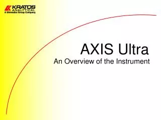
AXIS Ultra. An Overview of the Instrument. Ultra Spectrometer: Electron Optics. Analyser entrance slit plate. Lens V7. Electrostatic Lens. Lens V6. Lens V5. Lens V4. Apertures for definition of spot size. Lens V2. Scan Plates. Charge Neutraliser. Sample. Magnetic Lens.

E N D
AXIS Ultra An Overview of the Instrument
Ultra Spectrometer: Electron Optics Analyser entrance slit plate Lens V7 Electrostatic Lens Lens V6 Lens V5 Lens V4 Apertures for definition of spot size Lens V2 Scan Plates Charge Neutraliser Sample Magnetic Lens Magnetic immersion lens
Ultra Spectrometer: Spectral mode • Standard input lens optics is used • Electrons entering the analyzer slit with different energies (illustrated with different colours) are dispersed between the inner and the middle hemisphere • Common detector plane accomodates 8 MC detectors
Ultra Spectrometer: Imaging mode • Standard input lens is used • Third, “outer” hemisphere is added to create a spherical mirror analyzer (SMA) consisting of the middle and the outer spheres • Electrons pass through the middle hemisphere into the SMA & back to the detector plane • Parallel image maintained • Fast, real time image
The Spherical Mirror Analyser • First Investigated by Sar-El in 1966 and later by Tremblay and Roy 1983 and Diamon 1987 • Consists of two concentric hemispheres • Object and Image within inner hemisphere • Spherical aberration is zero • First order energy dispersion zero at image position Outer Hemisphere Innerhemisphere Object Image
Imaging properties of the spherical mirror • Left: Parallel electron trajectories at the SM analyzer entrance slit are parallel at the exit, therefore no first order radial dependence of the trajectory on the position in the image • Right: Electrons entering the analyzer at the same point with different energies will be dispersed within the SMA Energy dispersion Object Image Object Image
> Pass Energy Baffle Pass Energy < Pass Energy Object Energy Analysed Image Energy Analysis in the Spherical Mirror • Baffle with an aperture within the mirror field transmits only a small range of energies
XPS Imaging with the Kratos Ultra • Parallel images achieved in seconds • Variable FOV >2mm - 100mm • Spherical Mirror Analyser (SMA) works in FAT mode • Spatial resolution < 3mm
Examples:Cu grid • Cu grid • horizontal bars 20mm • vertical bars 40mm • Parallel images acquired in 1,5,10 & 20s • Fast photoelectron images allow sample alignment • As sample moves … image moves!!
XPS Imaging of 5 um Cu Grid • Example to follow! • 2 min acquisition shows excellent contrast • Retrospective line scans possible • gives 25mm pitch • Edge measurement (80-20%) • 2.2mm spatial resolution
XPS Imaging of 5 mm bars 2.2 um edge
XPS Imaging of Layered Materials • Survey of sample in cross-section showed Ti Al alloy Ti layer 8 um Al/SiC matrix
Ti Layer Summary • Ti 2p image acquired in 3 min! • Retrospective line scan shows ~10mm layer • Spectra acquired from 8mm layer • example of multipoint spectroscopy • good signal to background from < 10 mm area
What is multi-spectral imaging? • MCP and SMA used for spectroscopy! • FAT mode of SMA allows constant DE • Area limited by spatial resolution (~3mm) • Many areas from one image may be used
y(mm) Grey scale = intensity x (mm) Multi-spectral imaging Spectra reconstructed from small area eg (<10 mm) Energy (eV)
Spectra from Images • Record a sequence of images over any energy range • Sum intensity of pixels within defined area to obtain spectral info. • SiO/Si ring structure • 10m defined area in oxide and metal region • High resolution spectra
Example: Si/SiO2 • Patterned SiO2 on Si wafer • Multi-spectral imaging acquired through Si 2p peak • Images show complementary SiO2 and Si enriched areas • Si 2p spectra reconstructed from < 10 um areas • Note: Dark areas are covered by photoresist
Chemical state images SiO2 Si Si Decreasing BE SiO2
Spectra from Images Si oxide image Si metal image 10 um spot size
Example: Au dots on Si • 5mm Au dots on Si • Image acquired in 2 mins • Multi-spectral imaging performed through Au 4f7/2 peak • < 1eV resolution obtained from < 5mm!
Au 4f7/2 image and spectra Medium magnification Au 4f image acquired in 120 secs. Au 4f spectrum generated Au from 5mm area of image
Ag 3d3/2 with Spherical MirrorAnalyser Spectrum generated from parallel image using the Spherical Mirror Analyser. At 10eV pass energy, the Ag 3d5/2 spectrum generated using the SMA < 0.49eV FWHM At 20eV pass energy, the Ag 3d5/2 spectrum generated using the SMA < 0.49eV FWHM