Download
1 / 105
1.07k likes | 1.32k Vues
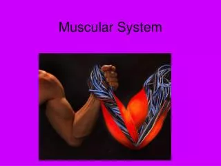
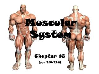
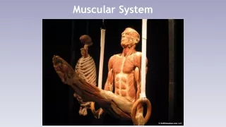
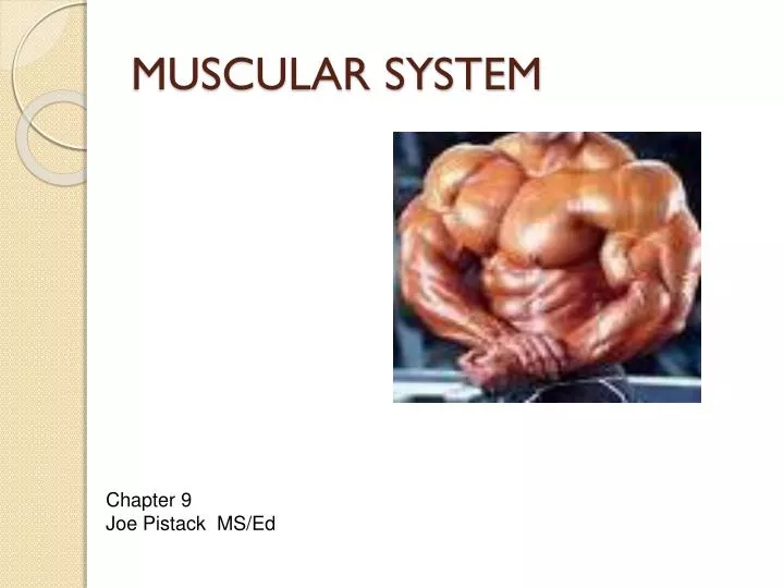
MUSCULAR SYSTEM. Chapter 9 Joe Pistack MS/Ed. MUSCULAR SYSTEM. The word muscle comes from the Latin word muse, which means little mouse . When a muscle contracts, the muscle movement under the skin resembles the movement of the mice scurrying around. TYPES OF MUSCLES.

E N D
MUSCULAR SYSTEM Chapter 9 Joe Pistack MS/Ed
MUSCULAR SYSTEM • The word muscle comes from the Latin word muse, which means little mouse. • When a muscle contracts, the muscle movement under the skin resembles the movement of the mice scurrying around.
TYPES OF MUSCLES Three types of muscle: Skeletal Smooth cardiac
SKELETAL MUSCLE -Generally attached to bone. -Voluntary muscle-controlled by choice. -Skeletal muscle cells are long, shaped like cylinders or tubes. -Appearance is striped or striated.
SKELETAL MUSCLE • Functions: • Produce movement. • Maintain body posture. • Stabilize joints. • Produce heat-helps to maintain body temperature.
SMOOTH MUSCLE • Generally found in the walls of the viscera. • Found in the bronchioles and blood vessels. • Involuntary-functions automatically. • Does not appear striped or striated. (non-striated)
CARDIAC MUSCLE • Cardiac Muscle-found only in the heart. • Function-pumps blood throughout the body. • Cardiac muscle is made of long branching cells that fit together tightly at junctions called intercalated discs. These discs promote the rapid conduction of electrical signals throughout the heart.
LAYERS OF CONNECTIVE TISSUE • Fascia-layers of tough connective tissue that surround large skeletal muscle. • Epimysium-the outer layer of the fascia. • Tendon-strong cordlike structure that extends toward and attaches to the bone.
LAYERS OF CONNECTIVE TISSUE • Perimysium-another layer of connective tissue, surrounds smaller bundles of muscle fibers. • Fascicles-bundles of muscle fibers. • Endomysium-third level of connective tissue the surround the individual muscle fibers.
COMPARTMENT SYNDROME • Compartment syndrome or crush syndrome, usually occurs from a crushing injury. • Ex. Pinned between two automobiles. • Usually occurs in the lower extremities. • A normal limb has an extensive amount of fascia that separates the muscle into isolated compartments.
COMPARTMENT SYNDROME • Each compartment receives the blood vessels and nerves necessary for muscle function. • In a “crush injury” the muscle is damaged, it becomes red and inflamed and leaks into the compartment. • Pressure in the compartment increases and compresses the nerves and blood vessels.
COMPRESSION SYNDROME • The muscles and nerves begin to die due to lack of oxygen and nourishment. • Immediate treatment involves reduction of the compartment pressure by slicing the fascia lengthwise.
MUSCLE ATTACHMENT • Muscles attach to other structures in three ways: • (1)tendon attaches the muscle to the bone. • (2)muscle attaches directly to the bone. • (3)sheet like fascia called aponeurosis connects muscle to muscle or muscle to bone.
STRUCTURE AND FUNCTION • Actin and Myosin are two proteins that are necessary for the contraction of muscles. • Sarcomere-contractile unit made up of myofibrils. • Muscles can only pull, not push. • When a muscle contracts it shortens.
HOW MUSCLES CONTRACT • Sliding filament theory-the interaction of actin and myosin sliding past each other causing the muscles to contract. • ATP and calcium play an important role in the contraction and relaxation of muscles.
SOMATIC MOTOR NEURON • Motor or somatic nerve-nerve that supplies skeletal muscle so that muscle contraction can take place. • A motor nerve comes from the spinal cord and supplies several muscle fibers with nerve stimulation. • Neuromuscular junction (NMJ)-area where the motor nerve meets the muscle.
MUSCLE STIMULATION • Electrical signal is given muscle membrane. • Triggers a series of events. • Results in muscle contraction.
DISORDERS OF NMJ • Myasthenia Gravis-disease that attacks the neuromuscular junction. • Symptoms-are due to damaged receptor sites on the muscle membrane. • Muscle contraction is impaired and the person experiences extreme weakness. • As the disease progresses, the person experiences difficulty breathing since breathing muscles are skeletal muscles.
TETANUS • Neurotoxins are transmitted by certain bacteria. • Clostridium tetani secretes a neurotoxin that causes excessive firing of the motor nerves. • Results in severs muscle spasm and tetanic contractions.
RESPONSE OF WHOLE MUSCLE • Single muscle fiber-contracts in an all-or-nothing response. • Whole muscle-capable of contracting partially, can contract weakly or very strong. • Ex. Small force is necessary to lift a pencil, greater force is necessary for 100lb. Weight.
RESPONSE OF A WHOLE MUSCLE • Recruitment-the process of recruiting additional fibers to achieve a greater muscle force. • Twitch-muscle contracts and then fully relaxes in response to a stimulus. • Tetanus-if a muscle is stimulated immediately and has no time to relax it remains contracted. Sustained muscle contraction is called tetanus.
MUSCLE TONE • Tetanic muscle contractions play an important role in maintaining posture. • The muscle being able to tetanize gives us an upright posture.
TONUS • Tonus-the normal continuous state of partial muscle contraction. • Muscle tone in the smooth muscle of the blood vessels helps to maintain blood pressure.
ENERGY SOURCE • Muscle contraction requires a rich source of ATP for energy, it is consumed by contracting muscles and replaced by: • (1)Aerobic metabolism • (2)Anaerobic metabolism • (3)Metabolism of creatine phosphate
ORIGIN AND INSERTION • Origin-attachment of the muscle to a stationary bone. • Insertion-attaches to a movable bone.
MUSCLE TERMS • Prime Mover-”chief muscle”, a single muscle that is generally responsible for most of the movement. • Synergists-”helper muscle”, works with other muscles. • Antagonists-muscles that oppose the action of other muscles.
MUSCLE TERMS • Hypertrophy-overuse of a muscle, the muscles increase in size.
ATROPHY • Atrophy-wasting away or decrease in the size of a muscle, due to lack of exercise or use.
CONTRACTURE • Contracture-abnormal formation of fibrous tissue within the muscle. • Result of the muscle being immobilized for long periods of time.
HOW MUSCLES ARE NAMED • The names of skeletal muscles are generally based on one or more of the following: • (1) size (5)number of origins • (2)Shape (6)origin & insertion • (3)direction of fibers (7)muscle action • (4)location
Muscles of the Head • Grouped into 2 categories: • (1) facial muscles • (2)chewing muscles • When facial muscles contract, they pull on the soft tissue. • This muscular activity is responsible for smiling and frowning.
Frontalis Muscle • Flat muscle-covers the frontal bone. • Extends from the cranial aponeurosis to the skin of the eyebrows. • Contraction of this muscle raises your eyebrows. • Gives us surprised look and wrinkles your forehead.
Orbicularis Oculi • Sphincter muscle that encircles the eye. • Sphincter-ring shaped muscle that controls the size of an opening. • Contraction of the muscle closes the eye and assists with winking, blinking and squinting.
Orbicularis Oris • Sphincter muscle that encircles the mouth. • Contraction of this muscle assists in closing the mouth, forming words, and pursing lips. • Kissing Muscle
Buccinator • Inserts into the orbicularis oris and flattens the cheek when contracted. • Used in whistling and playing the trumpet. • Considered the chewing muscle, on contraction it helps position food between the teeth for chewing.
The Zygomaticus • The smiling muscle. • Extends from the corners of the mouth to the cheekbones.
Chewing Muscles • Also called muscles of mastication(chewing). • Inserted on the mandible, or lower jaw bone. • Considered to be some of the strongest muscles in the body.
Masseter • Extends from the Zygomatic Process to the temporal bone in the skull to the mandible. • Contraction of this muscle closes the jaw. • Synergistically works with the other chewing muscles.
Temporalis • Fan-shaped muscle that extends from the flat portion of the temporal bone to the mandible. • Works synergistically with other chewing muscles.
Sternocleidomastoid • Contraction of only one of the sternocleidomastoid muscles causes the head to rotate toward the opposite direction. • Torticollis or wryneck is a spasm of this muscle. • Characterized by twisting of the neck and rotation of the head to one side.
Sternocleidomastoid • Extends from the sternum and clavicle to the mastoid process of the temporal bone in the skull. • Contraction of both muscles on either side of the neck causes flexion of the head. • The head bows as in prayer, called the Praying Muscle.