Download
1 / 29
530 likes | 1.36k Vues
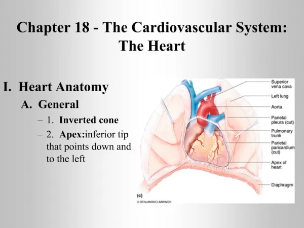
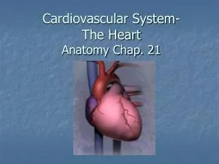
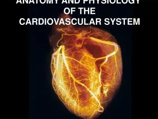
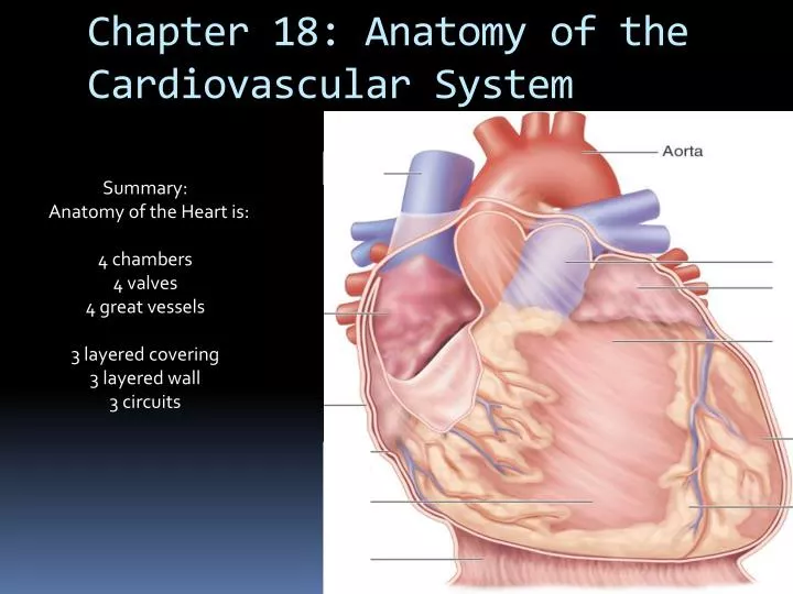
Chapter 18: Anatomy of the Cardiovascular System. Summary: Anatomy of the Heart is: 4 chambers 4 valves 4 great vessels 3 layered covering 3 layered wall 3 circuits. The Heart is a muscular PUMP (size and shape of a fist) with 4 Chambers

E N D
Chapter 18: Anatomy of the Cardiovascular System Summary: Anatomy of the Heart is: 4 chambers 4 valves 4 great vessels 3 layered covering 3 layered wall 3 circuits
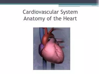
The Heart is a muscular PUMP (size and shape of a fist) with 4 Chambers The upper chambers ( R atrium, L atrium ) are for receiving Blood The lower chambers (R Ventricle, L Ventricle) are for pumping blood LA Right Atrium Left Atrium Right Ventricle Left Ventricle RA RV LV
Located in the MEDIASTINUM • of the THORACIC CAVITY, • between the Right and Left Lungs, • posterior to the body of STERNUM, • Anterior to Thoracic Vertebrae 5-8. • Sits atop the diaphragm • Right and Left Atria separated by the membranous ATRIAL SEPTUM (valves separate RA/RV, LA/LV) Ventricles separated by the muscular INTERVENTRICULAR SEPTUM RA LA --- -- --- -- -- - --- --- --- LV RV
4 CARDIAC VALVES • The Heart has 4valves, important in regulating the filling & flowing of the chambers of the blood TRICUSPID VALVE (Right Atrium / Right Ventricle) MITRAL VALVE (Left Atrium / Left Ventricle) PULMONIC VALVE (right Ventricle / Pulmonary trunk {artery} ) AORTIC VALVE (Left Ventricle / Aorta)
The Pulmonic and the Aortic Valves are semilunar valves, Each with three billowy, pocket-like leaflets The Atrioventricular valves (tricuspid and mitral) are composed of flat flaps (cusps), are connected to the interior ventricular walls via Connective tissue chords -- (CHORDAE TENDONAE), and PAPILLARY MUSCLES. Pulmonic valve Aortic valve LA RA Mitral valve LV Chordae tendonae Tricuspid valve RV Papillary muscle Chordae tendonae
Auricles of Right and Left Atria • On both the Right & the Left Atrium, • there is an ear-like extension called the Auricle these are visible on the external surface of the heart: Auricle of right atrium RA Auricle of right atrium RV Left ventricle Apex of the heart
The HEART and GREAT VESSELS • Each of the 4 cardiac chambers is associated with Major Blood vessel/s: (entering or exiting) • Right Atrium : (in) Superior & Inferior Vena Cavae • Right ventricle: Pulmonary Trunk (R & L pulm. arteries) (out) • Left Atrium: (in) Pulmonary veins • Left Ventricle: Aorta (out)
Pulmonary Trunk: R & L Pulmonary arteries Superior Vena Cava Aorta (Anterior view) Pulmonary veins Inferior Vena Cava (more on these to follow) Note: red oxygenated, blue unoxygenated blood
To remember great vessels: in order VC, AA, PT, PV vena cavae, aortic arch, pulmonary trunk, pulmonary veins
Triple layered Covering of the heart: PERICARDIAL SACHeart coverings protect against friction • Fibrous Pericardium: the thick, tough outer sac, which is lined By the • Serous Pericardium: a thin, moist double membrane, the parietal layer, which lines the fibrous pericardium, and the visceral layer, which adheres to & covers the heart, it is also known as THE EPICARDIUM
Coverings of the Heart • Fibrous pericardium • Serous pericardium (two-layered) • Parietal layer lines fibrous pericardium • Visceral layer (epicardium) forms outermost part of heart wall
3 Layers of the heart wall • Epicardium: thin , moist - is the visceral pericardial membrane • MYOCARDIUM - the Heart Muscle, the left ventricular wall is three times as thick as the right ventricle • ENDOCARDIUM - the inner lining, made up of single layer of ENDOTHELIUM, ( endothelium also lines the blood vessels of the entire CVS)
Ventricles ( FYI ) Two lower chambers known as pumping chambers because, upon contraction, they push blood into the large network of vessels Ventricular myocardium is thicker than the atrial myocardium because great force must be generated to pump the blood a large distance, against systemic resistance. Myocardium of left ventricle is thicker than the right for same reasons – distance and increased resistance.
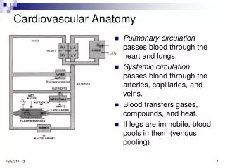
2 3 CIRCUITS • A. PULMONARY CIRCUIT: Right heart: unoxygenatedblood from SVC & IVC Right Atrium >>> Right Ventricle>>> Pulmonary Trunk Right and left Pulmonary arteries, into the LUNGS, for gases exchange; then to Heart via Pulm veins • B. SYSTEMIC CIRCUIT: Left heart: oxygenated blood from PULM Veins Left atrium >>> Left Ventricle >>> Aorta , and the systemic arterial, capillary, venous network C. CORONARY CIRCULATION: blood flow to the heart
***Flow of Blood Through Heart*** • Right side of heart is pulmonary circuit pump • Left side of heart is systemic circuit pump right atrium (tricuspid valve) -> right ventricle (pulmonary SL valve) ->lungs -> left atrium (bicuspid (mitral) valve) -> left ventricle (aortic SL valve) -> body tissues
Lung Bypasses in Fetal Heart • Foramen ovale– opening between right &left atria; after birth closes to form fossaovalis. • Ductusarteriosus– connection between pulmonary trunk & aorta; closes to form ligamentumarteriosum.
CORONARY ARTERIES:BLOOD SUPPLY TO THE HEART • THE FIRST BRANCHES off the AORTA, immediately superior to the Aortic valve: RIGHT CORONARY ARTERY, LEFT MAINSTEM CORONARY ARTERY, with its 2 quick branches: LEFT CIRCUMFLEX, LEFT ANTERIOR DESCENDING. Coronary blood flow actually occurs when the aortic valve cusps are closed, during back-flow; not during the powerful “systolic” pulsation of blood out of the ventricle during contraction.
(systole) (diastole)
CORONARY VEINS • Veins of the coronary circulation • As a rule, veins follow a course that closely parallels that of coronary arteries. After going through cardiac veins, blood enters the CORONARY SINUS to drain into the right atrium • Several veins drain directly into the right atrium
posterior anterior