Download
1 / 87
970 likes | 1.64k Vues
Upper & Lower Gastro-Intestinal Problems. Management of the Patient with Digestive Disorders. Conditions of the Upper GI Tract. GastroEsophageal Reflux Disease (G.E.R.D.) Gastritis Peptic Ulcer Disease (PUD) Gastric CA. CM’s & causes. Chemical irritation

E N D
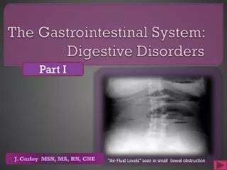
Upper & Lower Gastro-Intestinal Problems Management of the Patient with Digestive Disorders Marjorie Miller MA RN Timothy Frank MS RN
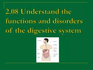
Conditions of the Upper GI Tract GastroEsophageal Reflux Disease (G.E.R.D.) Gastritis Peptic Ulcer Disease (PUD) Gastric CA
CM’s & causes Chemical irritation of nerve endings Pain or Tenderness Slow emptying (Gastric stasis) Anorexia or fullness Nausea Tension on walls • Medula stimulation • Nerve impulses: • (CTZ, GI, inner ear) Vomiting
CM’s & causes Local trauma or irritations to mucosa Bleeding • Peristalsis d/t • gastrocolic reflex • effort to rid toxin Diarrhea Belching & flatulence Swallowed air Incomplete digestion GI disease, gas forming foods, poor manners, food allergy Indigestion
Gastroesophageal Reflux Disease (GERD): Pathophysiology Length & frequency of esophageal acid exposure To HCl, Pepsin, bile acids & pancreatic enzymes pH < 2.0 Diffusion potential @ surface epithelial cells Cellular permeability H+ penetrate intracellular space H+ reach deeper sensory nerve endings “Heartburn”
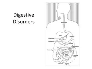
Sites of GI Pathology Esophogeal cancer Esophogeal Varices Gastritis Gastric Ulcer Gastric Cancer Duodenal Ulcer
ACUTE Gastritis • Transient inflammation of gastric mucosa • Common causes • Bacterial endotoxin (H.pylori, Staph) • Caffeine, Alcohol, Smoking • Steriods, Aspirin & NSAIDS • Bile Reflux • Burns, Shock, Sepsis • Severity • Moderate edema • Hemorrhagic erosion
Break in mucosal Barrier Vessel Erosion Acute Gastritis Pathophysiology Diffusion: HCl & Pepsinogen into mucosa Disruption of Mucosa & Capillary walls Loss of plasma proteins into gastric lumen Histamine Tissue Edema (increased capillary permeability & vasodilation) Prostaglandins Hemorrhage
Aspirin Unaware Heartburn Sour stomach Staphylococcus Abrupt & violent onset 5 hours after ingestion of contaminated food CM’s r/t cause Caffeine Tea Pepper Alcohol Transient vomiting GI bleeding Radiation Chemo Complete regeneration of mucosa within several days
Gastric Tissue Perfusion altered r/t blood loss nutritional 2° loss of acid-secreting cells Ineffective Breathing Pattern r/t pressure against diaphragm 2 GERD Abdominal distention & pain Gastritis –Nursing Diagnosis
Type A – fundal Autoimmune Circulating antibodies to Parietal cells Intrinsic factor Associated with Pernicious Anemia Addison’s Disease Hashimoto’s Thyroiditis Multiple bouts Type B – antral H. pylori Atrophy of gastric mucosa Chronic Gastritis - Causes
Pathophysiology - H. pylori & chronic gastritis/PUD • ½ World Population colonized/infected-but many do not develop disease. • H. Pylori bacteria: • duodenal bicarbonate secretion • contains proteases that degrade mucosa & • develops Peptic Ulcer disease; or, metaplastic ’s produces chronic gastritis gastric CA
First stage H. Pylori penetrates mucosal layer & forms clusters near membranes of surface epithelial cells
Second stage Some H. pylori attach to cell membrane Some lodge between epithelial cells
Diagnostic Studies for H. pylori • Noninvasive: • Stool or Breath Testing • Invasive: Biopsy of antral mucosa with rapid urease testing
Combination therapy can eradicate H. pylori in up to 85% of cases Antibiotics Antisecretory cytoprotective Amoxicillin Clarithromycin Flagyl Proton Pump Inhibitors Cytotec Carafate Pepto-Bismol H. Pylori – Medical Management Triple therapy for 7-14 days – Am GI Asso. Institute Review- 2008
Stress/Drug Related Mucosal Disease(SRMD)aka Peptic Ulcer Disease (PUD) Types/Cause: Duodenal Ulcers acid secretion Rapid gastric emptying buffering effect of acid load in duodenum penetrating lesion 1st 1-2 cm Gastric Ulcers Gastric erosion Break in mucosal barrier d/t incompetent Pylorus Superficial lesion Antrum
SRMD • Severe trauma • Burns – Curling’s Ulcer • Head Injuries – Cushing’s Ulcer • NSAID’s, aspirin, steroids, ETOH • Shock/Sepsis
Hemorrhage Prevalence 25% of clients have massive bleed– 2500 ml Onset Sudden Insidious Complications of Gastritis: Upper GI Bleed Severity • Arterial • Venous • Capillary
Significance of VS BP - 10 mmHg HR - 20 bpm reflects a blood loss of at least 1000cc = > 20% of total blood volume
Physiology ReviewNervous System Stressor Vasomotor Medullary Center
Physiology ReviewEndocrine System Decreased Cardiac Output and Hypotension Stressor Renin Angiotensin I Angiotensin II Adrenal Cortex Mineralcorticoids Aldosterone vasoconstriction Increased Na+ absorption Posterior Pituitary Antidiuretic Hormone (ADH) Increased H2O absorption Increased Blood Volume/Pressure Increased Venous Return =Increased Cardiac Output
1st Level Assessment (CM’s) Stage 2-Compensatory • O2P 20 bpm > baseline, bounding BP normal or systolic, diastolic RR rate & depth resp. alkalosis Skin pale, cool, delayed CR • Neuro restless, irritable, apprehensive oriented X3, Pupils-dilated & reactive • F/E slight in urine output, + Tilt Test thirsty
Sites of UGI Bleeds Esophogeal Varices Mallory-Weiss Syndrome Esophogeal cancer Gastritis Duodenal Ulcer Gastric Ulcer Gastric Cancer
Etiology – Esophageal Bleeds Mallory-Weiss Non-perforating tear of the gastric mucosa Exacerbated during vomiting Associated with alcohol use hiatal hernias gastritis esophagitis Esophageal and Gastric Varices d/t chronic liver disease (portal hypertension, causing pressure & dilation in esophageal veins)
Assessment – UGI Bleed History (Hx): Chief Complaint (CC) & History of Present Illness (HPI) precipitating or alleviating factors substance use or abuse vomiting stools diet history stress
Clinical Manifestations UGI Bleed • Pain • burning or cramping in mid-epigastric area • Nausea & possibly vomiting • Normal or bowel sounds • Hemorrhage or perforation may be first symptom
Hemorrhage • More common in duodenal vs. gastric • Common with varicies • Clinical manifestations • hematemesis • bright red or • “coffee ground” • stools: melena, maroon or burgundy • fluid volume deficit • H&H, BUN initially because of FV , but once volume status is corrected, both will go down. Positioning for safety
Fluid volume deficit Altered tissue perfusion The percent of blood loss correlates with CM’s: LOC, skin signs & capillary refill BP, HR UOP HemorrhageNursing Dx
Collaborative Management Hemorrhage • Establish IV route • replace with crystalloids or colloids • replace clotting factors • Monitor vital signs frequently • Gastric lavage • large bore NG tube • room temperature saline • Anticipate transfer to critical care • Hemodynamic monitoring • Diagnostic Endoscopy
Ulcers Therapeutic endoscopy using contact probes - heater, laser or electro or argon plasma coagulation to coagulate bleeder Varices sclerotherapy injection - agent injected into bleeder to sclerose vessel variceal band ligation - causes thrombosis and fibrosis of bleeder vasopressin +/- IV nitroglycerin Balloon tamponade Collaborative Management - cont’d
Pre-procedure NPO S.O. to Drive Pt. Home Remove dentures/bridges During Procedure Monitor VS for BZD OD Mazicon on hand Post-procedure Sims position Gag reflex Monitor for vagal response Monitor for perforation Procedure Conscious Sedation Anticholinergics Anesthetic Spray to back of throat Left lateral position Pictures Biopsy Esophagastroduodenoscopy(EGD) Nursing Care
PUD – complications & Indications for Surgery Massive hemorrhage unresponsive to fluid replacement and EGD procedures Gastric Outlet Syndrome Perforation
Gastric Outlet Obstruction Hypertrophy Swelling, scarring, spasm Most common in pyloric area Atony & dilation Projectile vomiting Duodenum Antrum Pylorus
Potential Complication of PUD: Perforation Clinical manifestations • sudden onset of severe upper abdominal pain • may have N&V • rigid, board-like abdomen • absent bowel sounds • shallow, rapid respirations • free air on abdominal x-ray
Surgical Interventions • Gastric closure - closes perforation • Vagotomy - acid secretion in stomach • Billroth I&II - vagotomy & antrectomy with anastamoses • Total gastrectomy - removes source of acid
Post-op Care NG tube management • patency, position (co2, pH paper) & stability observe, record and report output Fluid replacement • IV fluids • blood products Pain management • Cough, Deep Breathe, Ambulate Bright red/24 Dark red/ PO Day 1 Red/green PO Day 2 Bile color PO Day 3
Post-op Care • Post-op concerns: • Dumping syndrome • Post Prandial hypoglycemia • bile reflux gastritis
Dumping Syndrome – CM’s Food “dumps” into intestine Hyperosmolar bolus Rapidly pulls extracellular fluid into bowel Activates Sympathetic NS Fluid shift circulating blood volume • HR • Palpitations • Syncope • Skin signs • GI symptoms Distributive shock
Dumping Syndrome – CM’s Food “dumps” into intestine Hyperosmolar bolus Rapidly pulls extracellular fluid into bowel Activates Parasympathetic NS Distended bowel lumen • Distention • Cramping • Borborygmi • tenesmus
5-6 small meals fat, PRO, CHO roughage Liquids between meals only Develop a diet plan for the patient to prevent “dumping syndrome” based on the RX on the left side Management of Dumping Syndrome
Pt. - Family Education • risk factors • medication regime: sedatives & anticholinergics/antispasmotics to slow transit time • stools for occult blood • when to notify healthcare provider
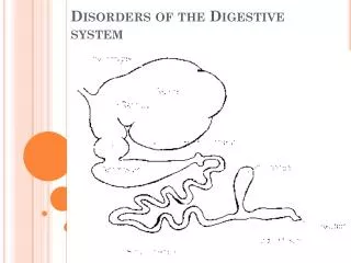
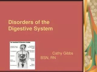
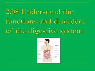
Conditions of the Lower GI (intestinal) tract • Peritonitis • Inflammatory Bowel Disease (IBD) • Crohn’s Disease • Ulcerative Colitis • CA colon
Peritonitis Inflammation of the large semi-permeable peritoneal double- layered membrane that covers the viscera and lines the walls of the abdominal and pelvic cavities
Peritonitis • Positive characteristics • exudes a thick, fibrinous substance in response to inflammation • adheres to other structures (mesentery & omentum) to “wall off” infection. • sympathetic stimulation gastric motility which inhibits spread of contaminants
Peritonitis • Negative characteristics • Large unbroken space: • Large surface area: favors transmission of contaminants permits rapid absorption of bacterial contaminants into the blood
Chemical gastric rupture ulcer ectopic bacterial Bacterial trauma ruptured peritoneal appendix dialysis pancreatitis Peritonitis – Types/Causes Beware: risk for peritonitis w/ruptured appendix
Pathophysiology Peritonitis Inflammation Shifts fluid volume from IVC to peritoneal space Peristalsis Free Air pressure fluid accumulation circulating volume O² requirements d/t pressure on diaphragm
Clinical Manifestations Peritonitis • Pain • Well localized Rigid abdominal muscles • with movement or pressure Guarding behavior • N & V F&E Imbalances • BS: Ø • Resp: shallow • WBC >20K CBC : Hb • grade fever <100-101F.