Download

1 / 35
420 likes | 775 Vues
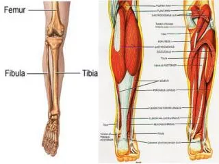
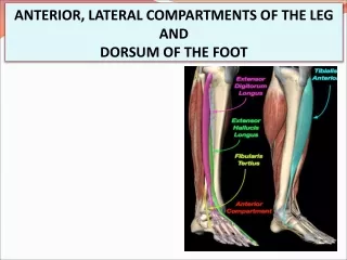
Fascial Compartments of the Leg. Dr. Zeenat Zaidi. Deep Fasia of the Leg. Continuous above with the deep fascia of the thigh (fascia lata) Below the tibial condyles, attached to the periosteum on the anterior and medial borders of the tibia

E N D
Fascial Compartments of the Leg Dr. Zeenat Zaidi
Deep Fasia of the Leg Continuous above with the deep fascia of the thigh (fascia lata) Below the tibial condyles, attached to the periosteum on the anterior and medial borders of the tibia Two intermuscular septae pass from its deep aspect to be attached to the fibula Together with the interosseous membrane, the septae divide the leg into anterior, lateral, posteriorcompartments Distally, the fascia thickens and forms the flexor, extensor, and fibular retinaculae
Retinacula • Around the ankle, the deep fascia thickens to form retinacula that keep the long tendons around the ankle joint in position and act as pulleys. • Superior Extensor Retinaculum • Attached to the distal ends of the anterior borders of the fibula and tibia • Inferior Extensor Retinaculum • Y-shaped band located in front of the ankle joint. Fibrous bands separate the tendons into compartments, each of which is lined by a synovial sheath.
Flexor Retinaculum • Extends from the medial malleolus downward and backward to be attached to the medial surface of the calcaneum. • Binds the tendons of the deep muscles of the back of the leg to the back of the medial malleolus as they pass forward to enter the sole. • The tendons lie in compartments, each of which is lined by a synovial sheath.
Superior Peroneal Retinaculum • Connects the lateral malleolus to the lateral surface of the calcaneum. It binds the tendons of the peroneus longus and brevis • Inferior Peroneal Retinaculum • Binds the tendons of the peroneus longus and brevis muscles to the lateral side of the calcaneum. The tendons each possess a synovial sheath, which is continuous above with the common sheath.
Muscles of the Leg: Movements The various leg muscles act on multiple joints and produce the following movements: Ankle:Dorsiflexion and Plantar flexion Intertarsal joints:Inversion and Eversion of the foot Toes:Flexion and Extension
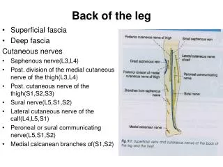
Upper part of the lateral surface: Lateral cutaneous nerve of the calf, a branch of the common peroneal nerve Lower part of the anterolateral surface: Superficial peroneal nerve, a branch of the common peroneal nerve Anteromedial surface of the leg: Saphenous nerve, a branch of the femoral nerve Cutaneous Nerves
Numerous small veins curve around the medial aspect of the leg and ultimately drain into the great saphenous vein The lymphatics follow the great saphenous vein, to end in the vertical group of superficial inguinal lymph nodes. A small amount of lymph from the upper lateral part of the front of the leg may pass via vessels that accompany the small saphenous vein and drain into the popliteal nodes Superficial Veins & Lymphatics
Muscles: Tibialis anterior Extensor digitorum longus Peroneus tertius Extensor hallucis longus Blood supply: Anterior tibial artery Nerve supply: Deep peroneal nerve Contents of the Anterior Compartment
Muscles of the Anterior Compartment • These musclesare the primary toe extensors and dorsiflexors. • Tibialis anterior plays an important role in holding up the medial longitudinal arch in the foot • Supplied by deep peroneal nerve
Origin: Lateral surface of shaft of tibia and interosseous membrane Insertion: Medial cuneiform and base of first metatarsal bone Muscles Origin: Anterior surface of shaft of fibula Insertion: Base of fifth metatarsal bone Origin:Anterior surface of shaft of fibula Insertion: Base of distal phalanx of great toe Origin: Anterior surface of shaft of fibula Insertion: Extensor expansion of lateral four toes
Blood Vessels Anterior Tibial Artery • Smaller of the terminal branches of the popliteal artery. • Arises at the level of the lower border of the popliteus muscle • Passes forward into the anterior compartment of the leg through an opening in the upper part of the interosseous membrane. • Descends on the anterior surface of the interosseous membrane,accompanied by the deep peroneal nerve.
In the upper part of its course: Lies deep to the muscles In the lower part of its course: Lies superficial in front of the lower end of the tibia. Passes deep to the extensor retinacula Lies between the the tendon of the extensor hallucislongus on its medial side and the deep peroneal nerve and the tendons of extensor digitorumlongus on its lateral side. It is here that its pulsations can easily be felt in the living subject. In front of the ankle joint, the artery becomes the dorsalispedis artery
Venaecommitantesof the anterior tibial artery join those of the posterior tibial artery in the poplitealfossa to form the popliteal vein Branches Muscular branches to neighboring muscles Anastomotic branches that anastomose with branches of other arteries around the knee and ankle joints
Nerve Supply Deep Peroneal Nerve • One of the terminal branches of the common peroneal nerve • Arises in the substance of the peroneus longus muscle on the lateral side of the neck of the fibula • Enters the anterior compartment by piercing the anterior fascial septum • Descends deep to the extensor digitorum longus muscle, first lying lateral, then anterior, and finally lateral to the anterior tibial artery • Passes deep to the extensor retinacula • Branches • Muscular • Articular branch to ankle joint
Contents of the Lateral Compartment • Muscles: • Peroneuslongus • Peroneusbrevis • Blood supply: • Branches from the peroneal artery • Nerve supply: • Superficial peroneal nerve
Muscles of the Lateral Compartment • The peroneus longus and brevis muscles are: • Planterflexors and evertors of the foot • Play an important role in holding up the lateral longitudinal arch in the foot. • In addition, the peroneus longus tendon serves as a tie to the transverse arch of the foot.
Origin: Lateral surface of shaft of fibula Insertion: Base of first metatarsal and the medial cuneiform Origin:Lateral surface of shaft of fibula Insertion: Base of fifth metatarsal bone
Artery of the Lateral Compartment • Numerous branches from the peroneal artery, which lies in the posterior compartment of the leg, pierce the posterior fascial septum and supply the peroneal muscles.
Nerve Supply Superficial Peroneal Nerve • One of the terminal branches of the common peroneal nerve • Arises in the substance of the peroneus longus muscle on the lateral side of the neck of the fibula • Descends between the peroneus longus and brevis muscles, and in the lower part of the leg it becomes cutaneous
Branches Muscular branches to the peroneus longus and brevis Cutaneous: Medial and lateral branches are distributed to the skin on the: Lower part of the front of the leg Dorsum of the foot Dorsal surfaces of the skin of all the toes, except the adjacent sides of the first and second toes and the lateral side of the little toe
The skin is thin, hairy, and freely mobile on the underlying tendons and bones. The sensory nerve supply is derived from the:. Superficial peroneal nerve supplies the skin on the dorsum of the foot; the medial side of the big toe; and the adjacent sides of the second, third, fourth, and fifth toes. Deep peroneal nerve supplies the skin of the adjacent sides of the big and second toes
The nail beds and the skin covering the dorsal surfaces of the terminal phalanges are supplied by the medial and lateral plantar nerves Saphenous nerve passes onto the dorsum of the foot in front of the medial malleolus. It supplies the skin along the medial side of the foot as far forward as the head of the first metatarsal bone. Sural nerve enters the foot behind the lateral malleolus and supplies the skin along the lateral margin of the foot and the lateral side of the little toe.
Dorsal Venous Arch • Lies in the subcutaneous tissue over the heads of the metatarsal bones • Drains on the medial side into the great saphenous vein and on the lateral side into the small saphenous vein. • The great saphenous vein leaves the dorsum of the foot by ascending into the leg in front of the medial malleolus. • The small saphenous vein ascends into the leg behind the lateral malleolus. • Tributaries: The greater part of the blood from the whole foot drains into the arch via digital veinsand communicating veins from the sole, which pass through the interosseous spaces.
Muscles of the Dorsum of the Foot Long Extensor Tendons • The tendon passes beneath the extensor retinacula, in company with the peroneus tertius muscle and then divides into four, which fan out over the dorsum of the foot and pass to the lateral four toes. • Opposite the metatarsophalangeal joints of the second, third, and fourth toes, each tendon is joined on its lateral side by a tendon of extensor digitorum brevis • On the dorsal surface of each toe, the extensor tendon joins the fascial expansion called the extensor expansion. Near the proximal interphalangeal joint, the extensor expansion splits into three parts: a central part, which is inserted into the base of the middle phalanx, and two lateral parts, which converge to be inserted into the base of the distal phalanx • The dorsal expansion, as in the fingers, receives the tendons of insertion of the interosseous and lumbrical muscles.
Extensor Digitorum Brevis Origin: Calcaneum Insertion: By four tendons into the proximal phalanx of big toe and through long extensor tendons to second, third, and fourth toes Action: Extends toes
Synovial Sheath of the Tendon of Extensor Digitorum Longus • The extensor digitorum longus and peroneus tertius tendons are surrounded by a common synovial sheath as they pass beneath the extensor retinacula. • The sheath extends proximally for a short distance above the malleoli and distally to the level of the base of the fifth metatarsal bone.
Artery of the Dorsum of the Foot • Dorsalis Pedis Artery • Begins in front of the ankle joint as a continuation of the anterior tibial artery. • Terminates by passing downward into the sole between the two heads of the first dorsal interosseous muscle, where it joins the lateral plantar artery and completes the plantar arch. • It is superficial in position and is crossed by the inferior extensor retinaculum and the first tendon of extensor digitorum brevis. • On its lateral side lie the terminal part of the deep peroneal nerve and the extensor digitorum longus tendons. • On the medial side lies the tendon of extensor hallucis longus . Its pulsations can easily be felt.
Branches Lateral tarsal artery, which crosses the dorsum of the foot just below the ankle joint Arcuate artery, which runs laterally under the extensor tendons opposite the bases of the metatarsal bones. It gives off metatarsal branches to the toes. First dorsal metatarsal artery, which supplies both sides of the big toe
Nerve Supply of the Dorsum of the Foot Deep Peroneal Nerve • Enters the dorsum of the foot by passing deep to the extensor retinacula on the lateral side of the dorsalis pedis artery. • Divides into medial and lateral branches. • Medial branch supplies the skin of the adjacent sides of the big and second toes. • Lateral branch supplies the extensor digitorum brevis muscle. • Both terminal branches give articular branches to the joints of the foot.