Download
1 / 19
541 likes | 2.12k Vues
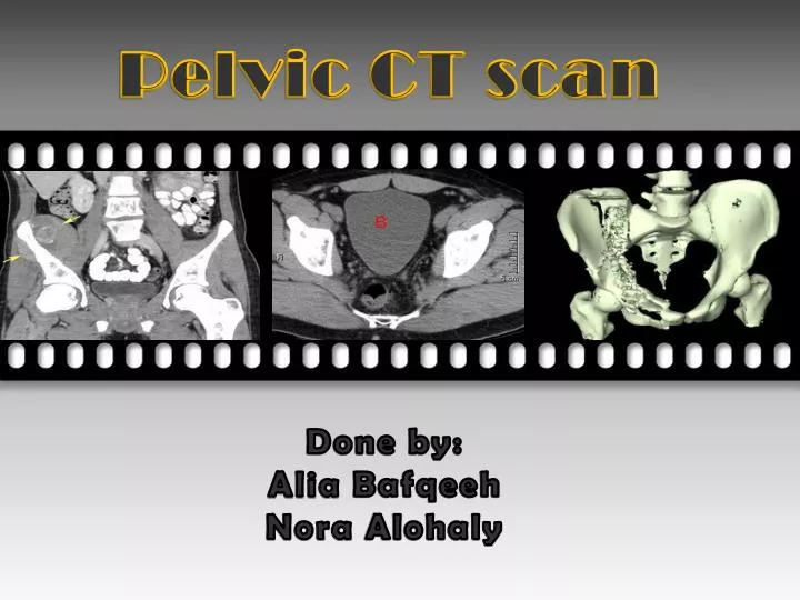
Pelvic CT scan. Done by: Alia Bafqeeh Nora Alohaly. Outlines :. Pelvic CT. Indications. Contraindications. Pelvic CT protocols. - Truma protocol. - Pathology protocol. Patient after care . . CT scan of the pelvis:.

E N D
Pelvic CT scan Done by: Alia Bafqeeh Nora Alohaly
Outlines : • Pelvic CT. • Indications. • Contraindications. • Pelvic CT protocols. • - Truma protocol. • - Pathology protocol. • Patient after care .
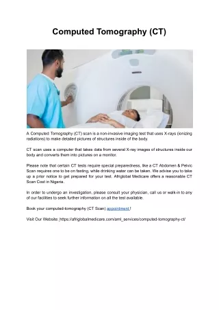
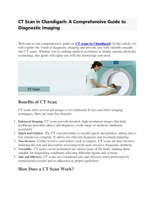
CT scan of the pelvis: It is an imaging method that uses x-rays to create cross-sectional pictures of the organs inside the pelvis (bladder, prostate, lymph nodes and pelvic bones).
Indications of pelvic CT : • Pelvic truma or fracture . • Hematuria or suspected renal calculus. • Hematoma. • Suspected hemorrhage. • Hip osteonecrosis. • Ischemic bowel. • Pelvic inflammatory or infection disease(abscess/colitis). • Pelvic vein thrombosis. • Congenital abnormalities e.g. CHD. • Tumors, suspected or known (Lymphadenopathy/Primary or metastatic malignancies) osteosarcoma of the ilium CHD Ewing sarcoma
Contraindications of pelvic CT : There are no absolute contraindications to pelvic CT examinations, the relative benefits should be outweigh the exposure risk. • Check the following conditions : • ALLERGIES, ASTHMA, DIABETIES, KIDNEY DISEASE • Ask if a patient is PREGNANT. • Ask about prior reaction to contrast.
Pelvic CT scan : • In KKUh there are 2 protocols • Truma . • Pathology .
Truma protocol (C-) E.g. to rule out fractures or history of truma . Patient preparation : No need for pt. preparation since he comes directly from the ER . - Pt. should be stable (vital sings). - On stretcher. • No need to be NPO except if a sedation is needed ( NPO for 3-4 hrs). Patient position : -Supine. -Feet first in gantry. -Hands above the head .
The vertical center is in the middle of the pelvis . • The axial center is in the iliac crest. • The scanning process : • Scout Images: • PA : plane 180º • Lat : plane 90º
The serial of procedure : Standard window Bone window
- When the scan is end we can have 2D reformats (coronal, sagittal ) and 3D pelvis .
pathology protocol (C+) E.g. Ca ovarian , mass or swelling Patient preparation : - 60 ml castor oil the night before the procedure. - NPO from mid night . - The patient should be in department 2hrs before start the procedure. - check that pt. not allergic or asthmatic . - Check pt. renal function test ; for inpatient 1 week outpatient (diabetic) 3 months outpatient (non-diabetic) 6 months - Pt. is given the oral contrast gastrografin or telebrix 3% (30 ml in 1000 ml of water ), one cup every 10 min .
After 1hr Pt. call and stay in beside room: • Pt. sign the consent form • Explain to patients the risks of contrast and answer any questions they have. • Check the blood pressure. • start Pt. cannulated with IV cannula (18-20 Gag). • The Pt. is then shift to the CT room . Patient position : -Supine. -Feet first in gantry. -Hands above the head . • The vertical center is in the middle of the body . • The axial center is in the xiphoid process. • NOTE: we must include the abdomen to avoid repeation & injection of CM twice .
The scanning process : • Scout Images: • PA : plane 180º • Lat : plane 90º • Inject the Pt. with IV contrast omnipaque or xenetix 300 by injector machine. • Flow rate 3ml/sec. • volume 120 ml. • By using the smart prep technique , after the CM is seen in the aorta in smart prep image the scan is delay for 65 sec ( portovenous phase ) .
When the scan is end we can have 2D reformats (coronal, sagittal ).
additional procedure : • The doctor may need • Delayed image after 10 min ( full KUB) to localized the area & size of the legion . • Rectal contrast 500 ml . showing a large filling defect (mass) extending posteriorly (arrows).
CT axial images with oral and intravenous contrast. A fluid filled diverticulum . (C) Coronal CT reformat shows fluid-filled diverticulum (arrow). (D) Sagittal CT reformat shows a diverticulum descending from the inferior border of the small bowel (arrow).
Patient after care : • The site of contrast injection will be bandaged. • The technologist will continue to watch the patient for possible adverse contrast reactions. • Pt. can eat or drink as normal. • - If the Pt. inject with contrast, he/she should drink plenty of liquid to help flush it out from there system.