Download
1 / 74
740 likes | 993 Vues
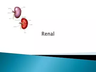
Renal Review. Ana Ivkovic and Rahul Dave. Kidney functions. Primary: water regulation and electrolyte balance-- homeostasis The renal system functions to maintain the intravascular volume (of body fluids) Other: Endocrine: renin, erythropoieten, calcitriol

E N D
Renal Review Ana Ivkovic and Rahul Dave
Kidney functions • Primary: water regulation and electrolyte balance--homeostasis • The renal system functions to maintain the intravascular volume (of body fluids) • Other: • Endocrine: renin, erythropoieten, calcitriol • Liver-like fxns: glucose synthesis
Basic Concepts • Excretion = Filtration - Reabsorption + Secretion • Filtration: Bowman’s capsule • Reabsorption: Peritubular capillaries • Secretion: Peritubular capillaries
Measuring Fluid Compartments • Total Body Water: varies with fat • 20-40-60 Rule • ICF high in K and Mg; ECF high in Na, Cl • Plasma high in protein; interstitial fluid low in protein-- Gibbs Donnan (neg. charged prots attract more pos. charge) • Smallest compartment (plasma) most important (intravascular volume that is controlled by kidney)
Osmolarity and Oncotic Pressure • Normal plasma osmolarity = 285-290 mOsm/L • Tightly controlled • Osmolarity vs. Osmolality • Osmolarity = mmol solute/L solution • Osmolality = mmol solute/kg h2O • Reflection coefficient: • 0 = ineffective osmolyte (urea, ethanol--freely permeable) • 1 = effective osmolyte (Na, K, glucose w/o insulin; “draw” water) • Oncotic Pressure: the fraction of plasma osmolarity that is due to plasma proteins
Tonicity vs. Osmolarity • Osmolarity • Describes the osmotic properties of a solution • Tonicity • Refers to the osmotic effect on the volume of a cell • Ex: hypotonic soln--water moves in, cell swell • Isosmotic solns not necessarily isotonic (has to do w/ concept of reflection coefficient--ex of urea solution and RBC)
Darrow-Yanet Diagrams--Think Logically! • All volume disturbances originate in the ECF compartment • Changes in the ICF compartment are in response to changes in the ECF • “hyposmotic contraction” refers to the volume of fluid that remains
Volume contractions • Diarrhea, vomiting, loss of blood--isosmotic volume contraction • Diaphoresis (sweating), dehydration--hyperosmotic contraction • Remember that sweat is hyposmotic • Addison’s disease (lack of aldosterone)--hyposmotic volume contraction
Volume expansions (rarer) • Isotonic volume expansion (isotonic saline IV): ECF expands, ICF doesn’t change • Hypertonic volume expansion: ECF osmolarity increases, draws fluid from ICF • Hypotonic volume expansion: ECF osmolarity decreases, adds fluid to ICF (examples: psychogenic polydipsia, SIADH)
Renal vascularization • Renal artery --> interlobar artery --> arcuate artery --> interlobular artery--> afferent arteriole* --> glomerular capillaries--> efferent arteriole* --> peritubular capillaries • *serial arrangement of arterioles--important!
Juxtamedullary vs. Superficial Nephron • JMN has long Loop of Henle • Generates a concentrated urine • JMNs are what we lose with age
Renal Clearance and Blood Flow • C.O. = 5.2 L/min • RBF = 1.2 L/min (20% of cardiac output) • RPF = .66 L/min (plasma = 55% of blood); also equal to the clearance of PAH (filtered and secreted) • GFR = Clearance of inulin or creatinine • Inulin is filtered but not secreted or reabsorbed • Creatinine clearance a slight overestimate of GFR because it is partly secreted (GFR = 0.9 X Ccreatinine) • Filtration Fraction = GFR/RPF, normally 20%
PAH • Used to measure RPF • Effective RPF = ([U]PAH x V) / [P]PAH = CPAH
Clearance Ratio • CR = Cx/Cin • If CR = 1, substance “x” is only being filtered • If CR < 1, substance “x” is being reabsorbed • If CR > 1, substance “x” is being secreted
GFR: • Is dependent on hydrostatic pressure inside glomerular capillaries • Depends on the oncotic pressure inside glomerular capillaries • Is equal to the clearance of inulin • Under normal conditions, is rarely dependent on the oncotic pressure inside Bowman’s space • Creatinine is used to calculate it • Three of the above • All of the above
Starling’s Forces of capillary exchange • GFR = Kf (PGC - PBS - ∏GC) • Hypoalbuminemia increases GFR • PBS: low unless obstruction present (kidney stones increase GFR) • Basement membrane has fixed negative charge--> neg. charged prots can’t get across --> oncotic pressure in Bowman’s space = 0
Cont’d • Hydrostatic Pressure is high and relatively constant (due to serial arterioles) • Oncotic Pressure increases along length of glomerular capillary (as more fluid is filtered out) • Filtration occurs upstream while reabsorption occurs downstream • Q: why does a low GFR result in increased reabsorption? • A: more time to filter --> oncotic pressure increases
AFFERENT AND EFFERENT ARTERIOLES ARE THE MAJOR SITES OF REGULATED RESISTANCE IN THE RENAL VASCULATURE: Glomerular capillary is unique: 2 sites of vasoconstriction
Autoregulation • Myogenic Mechanism (Bayless): intrinsic reflex mechanism of smooth muscle; increased pressure causes vasoconstriction • Tubuloglomerular feedback: macula densa senses increased filtered load of NaCl--> sends signals to afferent arteriole to vasoconstrict, thereby decreasing the filtered load (by decreasing GFR back to normal) • Both processes serve to keep RBF and GFR constant
Sympathetic Innervation • There is no parasympathetic input to the kidneys • Sympathetic innervation of the afferent and efferent arterioles is the major regulator of RBF and GFR • Vasoactive compounds also act on afferent and efferent arterioles: NE, Angiotensin II, Endothelin--> constrict; Ach, NO, PGs, etc --> dilate • Low vs. severe sympathetic drive--examples of exercise and hemorrhage
Urine formation • Ultrafiltration of plasma • Reabsorption of H2O and solutes from tubular fluid • Active and passive processes • Transcellular and paracellular (lateral space) transport; latter occurs in proximal tubule due to leaky tight junctions--> ions pass, followed by H2O • In collecting duct tight jxns are very tight and do not allow passage of water, proteins, or solutes
Reabsorption and Secretion along Proximal Tubule • Isosmotic fluid reabsorption • Reabsorbs 2/3 of filtered load of Na and water (Aquaporin 1) • Highly permeable to H2O; solvent drag of K and Ca • Understand TF/P graph
Upper Segment of PT • Na cotransported along with bicarb, glc, amino acids, phosphate (luminal membrane) • H+ secreted as counter-transport with Na (luminal membrane) • Sodium bicarbonate is reabsorbed (basolateral membrane) • Under normal conditions, reabsorption will increase as plasma [gluc] increases • Once plasma [gluc] reaches a certain level, all glucose carriers in the PT will be saturated, leaving some glucose behind • Tm of SGLT-2 (sodium coupled) is 200g/dl, which is exceeded in diabetics; osmotic diuresis results
Lower Segment of PT • NaCl reabsorbed transcellularly (1/3) and paracellularly (2/3); due to transepithelial voltage • Amino Acids and Bicarbonate have been completely reabsorbed • Glucose SGLT-1 (2 Sodium coupled) transporters move glucose against higher gradient
Thick Ascending Limb • Reabsorbs 1/4 of filtered Na • Has the Na-K-2Cl cotransporter • Inhibited by Furosemide (loop diuretic) • Impermeable to water; tubular osmolarity decreases (“diluting segment”)--> separation of movement of water and solute • Lumen becomes positively charged, causing paracellular transport of Na, K, Ca, and Mg
Early Distal Tubule/Collecting Duct • Also impermeable to water (like TAL) • Continues the dilution of urine; the “cortical diluting segment” • Reabsorption of Na/Cl (cotransporter) • Inhibited by Thiazide diuretics • Thiazide diuretics unique in that they increase Ca++ reabsorption (Loop diuretics increase Ca++ excretion by diminishing NaK2Cl + lumen effect)
Late Distal Tubule/Collecting Duct: fine tuning • Principal cells--reabsorb Na, H2O, and secrete K+ • Impermeable to water, except in presence of ADH (Vasopressin) • ADH causes water channels to relocate to apical cell membrane (AQUAPORIN 2) • Na (transcellularly) and Cl (paracellularly) are reabsorbed • Aldosterone causes an increase in Na absorption and increases K secretion • Spironolactone (K-sparing) blocks aldosterone; other K-sparing diuretics (Triamterene, Amiloride) act directly on the Na channel, independent of aldosterone • Intercalated cells--secrete H+ through primary active transport • exchange H+ out of cell for K+ into cell; K+ reabsorption • possess carbonic anhydrase activity for bicarb reabsorption
Miscellaneous Renal Stuff • Na+, Ca++ are never “secreted”;rather, fail to reabsorb • Prostaglandins released during hypovolemic shock to increase RPF and prevent renal ischemia • Aldosterone: promotes Na reabsorption and K secretion (via action on principal cells); also promotes H+ secretion (via action on intercalated cells)-->a link between volume and acid-base regulation • Posm = 2[Na] + 2[Glucose] + [BUN] • ADH: OSMOREGULATION • ALDOSTERONE: Na+/VOLUME REGULATION
Genetic Defects that Target Tenal Transport Mechanisms • Bartter’s Syndrome: defect in NaK2Cl transporter • Gettelman’s Syndrome: defect in Na/Cl cotransporter • Liddel’s: defect in ENaC (turned on) • Pseudohypoaldosteronism: defect in ENaC (turned off--> Na doesn’t get reabsorbed--> volume contraction
Urine flow rate is never zero • There is an inverse relationship between urine flow and osmolarity • Normal urine osmolarity ≈ 600 mOsm/L • Range = 50 - 1200!! (kidneys can concentrate urine up to 4x the plasma concentration)
Control Mechanisms of Osmoregulation • Osmoreceptors • Increase in plasma osm--> hypothalamus stimulated to release ADH (hypothalamic set point ≈ 285 mOsm/L solution) • Respond to < 2% change in plasma osmolarity • Most important control in osmoregulation • Baroreceptors • Respond to changes in Blood Pressure • Require a 15-20% change in BP before activation
Disorders of Osmoregulation • Psychogenic Polydipsia, Hypothalamic/Central Diabetes Insipidus: low ADH • Nephrogenic Diabetes Insipidus: ineffective ADH (kidney unable to respond to ADH)
Mechanisms to Concentrate Urine • Countercurrent Multiplication--creation of osmotic gradient • Loop of Henle • Generates a urine that is concentrated as high as 600 mosm/L • Urea recycling • Medullary Collecting Duct • Needed to increase the osmolar gradient from 600 to 1200 mosm/L • Kidneys use urea to do osmotic work when in state of antidiuresis • Countercurrent exchange--vasa recta maintains the medullary insterstitial osmotic gradient set up by the countercurrent multiplier
Diuresis vs Antidiuresis • Understand the diagrams on p. 9 • Water diuresis: most concentrated urine just before ascending limb and TAL; most dilute at end of CD • Antidiuresis: most concentrated in lumen at level of renal papillae (in medulla); most dilute at TAL
Renin, Angiotensin, Aldosterone • Renin secretion stimulated by: • Decrease in effective circulating volume (decreased pressure at afferent arteriole) • Increase in sympathetic nerve activity • Tubuloglomerular feedback (decreased Na load sensed by macula densa, causing renin release) • Angiotensin II: • Arteriolar constriction--> increases TPR • Increases Aldosterone • Increases ADH and thirst • Aldosterone causes: • Na reabsorption at principal cells • K secretion in CD
Aldosterone secretion: • Increased by ACTH, Angiotensin II, high plasma [K+], cases of volume contraction • Decreased by *ANP* and high plasma Na+ (feedback inhibition) • ANP: OPPOSES RAAS; increases Na+ excretion during cases of volume expansion (cardiac myocytes are stretched) Note: Na+ alterations do not affect plasma osmolarity, rather they affect the effective circulating volume; H2O homeostasis and ADH determine plasma osmolarity
Aldosterone escape A protective mechanism during cases of abnormal aldosterone elevation (example of adrenal tumor); system becomes insensitive to aldosterone.
Renal Physiology Lectures 41 to 48 Rahul Dave’ (rdave2@uic.edu)
I can’t go through everything in detail. Know the handout. My goal is to make this make sense to you, and orient your studying. Pay attention to major vs minor factors. Minor doesn’t mean less important to study, but helps you keep changes in perspective You need to memorize the regulation, etc … and understand the logic. It’s easy to talk yourself into something wrong.
K+ distribution and homeostasis • Plasma K is low … must be controlled well • Determines membrane potential • Metabolic alkalosis causes hypokalemia (and vice-versa) • Rules of thumb: understand mechanisms • Na and K go opposite
K+ Transport K+ Transport See diagrams in handout for cellular transport pathways in different sections
Regulation of K+ MAJOR FACTORS K+ itself (K promotes its own secretion) Aldosterone (Na+ excr., K+ reabs., H+ excr.) MINOR FACTORS Tubular flow increases secretion ADH no net effect Alkalosis (acute) increases secretion Tubular Na+ increases secretion Insulin Increase reabsorption Epinephrine Decreases secretion (fight/flight)
Diuretics See diagrams of cell transport pathways