Download
1 / 19
190 likes | 211 Vues
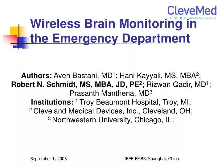
Wireless Brain Monitoring in the Emergency Department. Authors: Aveh Bastani, MD 1 ; Hani Kayyali, MS, MBA 2 ; Robert N. Schmidt, MS, MBA, JD, PE 2 ; Rizwan Qadir, MD 1 ; Prasanth Manthena, MD 3 Institutions: 1 Troy Beaumont Hospital, Troy, MI;

E N D
Wireless Brain Monitoring in the Emergency Department Authors: Aveh Bastani, MD1; Hani Kayyali, MS, MBA2; Robert N. Schmidt, MS, MBA, JD, PE2; Rizwan Qadir, MD1; Prasanth Manthena, MD3 Institutions:1 Troy Beaumont Hospital, Troy, MI; 2 Cleveland Medical Devices, Inc., Cleveland, OH; 3 Northwestern University, Chicago, IL; IEEE-EMBS, Shanghai, China
Objective To evaluate the feasibility, quality and utility of four-channels of electroencephalogram (EEG) telemetered from patients presenting with Altered Metal Status in the Emergency Department. IEEE-EMBS, Shanghai, China
Background • Many modalities exist to evaluate neurologic status of patients such as: • Bispectral Index (BIS), • Cerebral Oximetry, or • Positron Emission Tomography (PET). • Despite some optimistic trials in the Emergency Department (ED) to evaluate the neurologic status of patients, none has supplanted the need for the Electroencephalogram (EEG). • EEG is the gold-standard for objectively evaluating the functional neurologic status of patients. IEEE-EMBS, Shanghai, China
Background - Continued • Only a handful of hospitals in the US routinely perform EEGs in the ED. Subsequently, patients in whom an EEG is required are admitted to the hospital with their potential disorder undiagnosed and untreated for many hours or days. • This Altered Mental Status group currently makes up 10% or 14 million of the 140 million yearly ED visits in the U.S. IEEE-EMBS, Shanghai, China
Background - Continued • Most emergency departments (ED) do not perform electroencephalogram (EEG) studies. This is due to several inhibiting factors including: • The bulk of the equipment makes it inconvenient to be permanently located in an ED setting, • The cost of equipment at $20,000-$40,000 per unit is very expensive for most ED budgets, • The time and expertise required to set up and monitor an EEG is typically lacking in the ED, and • ED personnel are not trained to read EEGs and a neurologist may not be immediately available to read the EEG. IEEE-EMBS, Shanghai, China
New EEG Equipment is Needed for the Emergency Department • The Crystal Monitor was developed under US National Institutes of Health Grants to have an ED EEG device • Wireless, small, unobtrusive • Low Cost, ~ 1/2-1/3 of most EEG machines • Fast, easy to use, limited number of channels • Easy to learn how to use IEEE-EMBS, Shanghai, China
The Study Guided by NIH recommendations and support, Cleveland Medical Devices Inc. (CleveMed) has created a portable telemetry multi-channel EEG monitor. A four-channel montage (Fp1-C3, Fp2-C4, C3-O1, C4-O2, Gnd FpZ) was used to maximize EEG coverage while minimizing electrode set-up time. Telemetry allows the patient to be un-tethered and moved about freely while still being monitored, an important requirement for any patient being assessed in the ED. An internet connection allows a neurologist to interpret the EEG from anywhere. IEEE-EMBS, Shanghai, China
Patient with Crystal Monitor 16 (older version) IEEE-EMBS, Shanghai, China
Inclusion Criteria: • The following patients will be eligible for study inclusion: • Patients with known seizure disorder of any type, but with prolonged (> 1 hr.) post-ictal mental status change. • Patients with status epilepticus who have received a muscle relaxant for intubation to determine the presence of subclinical seizures. • Patients with brief alteration of mental status of unknown origin. (This group includes new onset seizure disorder, syncope, “spells,” “blackouts,” etc.) • Patients with behavioral changes that may indicate nonconvulsive seizures (impaired consciousness, violent outbursts, unusual behaviors, etc.) • Acute head injury patients with mental status changes that may indicate nonconvulsive seizures. • Patients with a history of previous head injury presenting with new onset mental status changes. (Head injured patients are at risk for post-traumatic seizures). • Patients with neurological exams that may be consistent with focal or partial nonconvulsive seizure. (Eg: aphasia, Todd’s paralysis, etc.) IEEE-EMBS, Shanghai, China
Exclusion Criteria: • The following patients will be excluded from the above groups. • Patients who are convulsing. • Medically or surgically unstable patients. • Family member, other authorized representative unable to give informed consent. • Patients with a head injury incompatible with the use of EEG (eg: gunshots, severe scalp abrasions, etc.) IEEE-EMBS, Shanghai, China
Transmission and Reception: After EEG was completed, the data was password encrypted and transmitted to one of two study neurologists. • The neurologist would then provide a real-time read for the EEG via telephone conversation or email. • The neurologist also subjectively evaluated the quality of the EEG utilizing the following four point scale: ● 4 = Excellent quality/Acceptable ● 3 = Good quality/Acceptable ● 2 = Fair quality/Acceptable ● 1 = Poor quality/Unacceptable • Patients were followed to either attain their discharge diagnosis from the ED or the hospital in the case of admission. IEEE-EMBS, Shanghai, China
48.6 % of the Subjects had EEG’s that were Abnormal EEG interpretations (5 unusable EEG’s were not included): • 37/72 (51.4%) EEG’s were interpreted as normal • 2/37 were diagnosed as pseudoseizure by the ED physician • 28/72 (38.9%) EEG’s were interpreted as slowing • 11/28 were patients who clinically appeared post-ictal • 7/72 (9.7%) EEG’s identified a sub clinical epileptogenic foci Correlation with Standard Inpatient EEG: • 24/77 (31.1%) patients with EEG had an inpatient EEG • 18/24 (75%) were equivalent to the study EEG • The six dissimilar results are described below IEEE-EMBS, Shanghai, China
75% of the EEG’s Corresponded with the 32 Channel Clinical EEG Six ED EEG’s were different than the EEG Lab results that were performed later IEEE-EMBS, Shanghai, China
Discussion and Future Considerations Understanding that EEG is a time-sensitive modality, it is important that we perform EEG’s when they can be most useful, i.e. in the acute setting. No enrolled patient failed to complete an EEG. Only 5 of 77 patients (6.5%) had unusable EEG’s primarily due to combination of muscular artifact and gaps in the data for interference during wireless transmission. An improved radio that can re-transmit lost packets has been developed (Crystal Monitor Model 20) and will be used for the second half of this study. Based on this data we believe that ED EEG provides valuable information to the ED physician, which can expedite safe medical care. We do not assert that a four-channel EEG is superior or equivalent to the standard EEG. We do believe its use as a screening tool in the ED provides the ED physician with the additional information necessary to make a more appropriate disposition from the ED. IEEE-EMBS, Shanghai, China
Crystal Monitor 20 Specifications • Dimensions: 135 mm x 63 mm x 25 mm (5.3” x 2.5” x 1”) (not including antenna) • Weight: 210 grams (6.4 oz.) with batteries • Antenna: 76 mm (3.0“) flexible • Number of Input Channels: • 8 configurable channels (external sensors) plus: • 1 internal position sensor, • 1 pulse oximeter, • 1 airflow sensor, • 1 DC channel Input Range ± 750µV to ± 2V (configurable) • Resolution 8, 12, 16 bits, configurable • Sampling Rate 960 Samples per second per channel • Filter Input bandwidth 0.5 Hz - 250 Hz (-3dB attenuation); CMRR 100 dB • Noise < 2 µV peak-to-peak (0.5 Hz – 100 Hz) • Input Impedance > 20 MΩ @ 10 Hz • Input Interface Standard no-touch 1.5 mm connectors • Power Supply 2 AA alkaline batteries, Battery Life 12 hours continuous use IEEE-EMBS, Shanghai, China
Conclusions Four-channel telemetry EEG used in the ED is feasible, provides good quality screening EEG’s and was able to diagnose underlying seizure in a significant number of patients. IEEE-EMBS, Shanghai, China
ACKNOWLEDGMENT This work was supported by NIH Phase II SBIR Grant No. 5 R44 NS042977-03 National Institute of Neurological Disorders and Stroke US National Institutes of Health IEEE-EMBS, Shanghai, China
For Questions Contact Robert N. Schmidt Cleveland Medical Devices Inc. 4415 Euclid Ave., Suite 400 Cleveland, Ohio 44103 USA rschmidt@CleveMed.com Phone: 1-877-CleveMed (253-8363) (US Toll Free) Phone Direct: +01-216-619-5925 Fax: 216-791-6744 IEEE-EMBS, Shanghai, China
Smaller Hardware • Non-Programmable (factory settings) • 2 channels • 960 sps • Up to 12 bits • Input Selections • EEG +/- 1 mV, 0.1-70 Hz. • EKG +/- 5 mV, 0.1-150 Hz. • EMG +/- 50 mV, 0.1-500 Hz. • Range, 50 ft. • Low Noise , < 1 uV RMS • Low weight, 191 grams (0.42 oz.) • Battery options, 14 hrs to days BioRadio Jr. IEEE-EMBS, Shanghai, China