Download
1 / 31
390 likes | 886 Vues
Names of Muscles are Descriptive. Relative size Pectoralis major: major = large size Shape Deltoid: like a delta or triangle Location Extensor digitorum : digits = fingers or toes Action Extensor digitorum : extension Number of attachments Biceps brachii : biceps = 2 heads

E N D
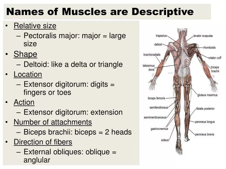
Names of Muscles are Descriptive • Relative size • Pectoralis major: major = large size • Shape • Deltoid: like a delta or triangle • Location • Extensor digitorum: digits = fingers or toes • Action • Extensor digitorum: extension • Number of attachments • Biceps brachii: biceps = 2 heads • Direction of fibers • External obliques: oblique = anglular
Muscle Facts • The human body has over 600 distinct skeletal muscles • The face contains over 60 muscles • 40 of which are used to frown • 20 of which are used to smile • The stapedius in the middle ear is the smallest muscle in the body • The gluteus maximus in the buttock is the largest muscle in the body • The sartorius in the thigh is the longest muscle in the body
Temporomandibular Joint (TMJ) • Articulation between the mandibular condyle of the mandible and the mandibular fossa of the temporal bone • Strain of the joint may be caused by • Grinding the teeth • Hyperextending the lower jaw • The condition of having strained the joint is called temporomandibular joint syndrome or TMJ syndrome • Symptoms include headache, earache, and pain in the jaw, neck, or shoulder
Muscles of Facial Expression (p. 195) • Enable communication through facial expressions of surprise, sadness, anger, fear, disgust, and pain • Examples: • Epicranius – lifts eyebrow • Made up of the frontalis & occipitalis • Orbicularis oris – closes lips • Orbicularis oculi – closes eyes • Buccinator – compresses cheeks • Zygomaticus – raises corner of mouth • Platysma – draws angle of mouth downward
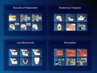
Muscles of Mastication (p. 195) • Enable chewing movements • Three pairs of these muscles are used in biting movements: • Masseter – elevates the mandible • Temporalis – elevates the mandible • Sphenomandibularis • Newly discovered muscle • Extends about an inch and a half from behind the eyes to the inside of the jawbone • Thought to help produce the movements of chewing
Muscles That Move the Head (p. 196) • Result from the actions of paired muscles in the neck and upper back • Examples: • Sternocleidomastoid • Pulls head to one side • Pulls head toward chest • Raises sternum • Splenius capitis • Rotates head • Bends head to one side • Brings head upright • Semispinaliscapitis • Extends head • Bends head to one side • Rotates head
Muscles That Move the Pectoral Girdle • See page 196 • Closely associated with muscles that move the arm • Examples: • Trapezius • Rotates scapula & raises arm • Raises scapula • Pulls scapula medially • Pulls scapula and shoulder downward • Rhomboid major • Raises and adducts scapula • Levator scapulae • Elevates scapula
Muscles That Move the Pectoral Girdle • See page 196 • Continued • Serratus anterior • Pulls scapula anteriorly and downward • Pectoralis minor • Pulls scapula anteriorly and downward • Raises ribs
Muscles That Move the Arm (p. 198) • The arm is one of the more freely movable parts of the body • Muscles connect the humerus to many other parts including the pectoral girdle, ribs, and vertebral column • Examples: • Coracobrachialis • Flexes and adducts arm • Pectoralis major • Pulls arm anteriorly and across chest • Rotates humerus • Adducts arm
Muscles That Move the Arm (p. 198) • Arm muscles (continued) • Teres major • Extends humerus • Adducts and rotates arm medially • Latissimusdorsi • Extends and adducts arm • Rotates humerus inwardly • Pulls shoulder downward and posteriorly • Supraspinatus • Abducts arm • Deltoid • Abducts arm, • Extends or flexes humerus
Muscles That Move the Arm (p. 198) • Arm muscles (continued) • Subscapularis • Rotates arm medially • Infraspinatus • Rotates arm laterally • Teres minor • Rotates arm laterally
Muscles That Move the Forearm (p. 199) • Muscles that connect the radius or ulna to the humerus or pectoral girdle produce most of the forearm movements • Examples: • Biceps brachii • Flexes forearm at the elbow • Rotates hand laterally • Brachialis • Flexes forearm at elbow • Brachioradialis • Flexes forearm at elbow • Triceps brachii • Extends forearm at elbow
Muscles That Move the Forearm (p. 199) • Forearm muscles (continued) • Examples: • Supinator • Rotates forearm at elbow • Pronator teres • Rotates forearm medially • Pronator quadratus • Rotates forearm medially
Muscles That Move the Hand (p. 200) • Muscles that move the hand originate from the distal end of the humerus and from the radius and ulna • Two major groups: • Flexors – anterior forearm • Extensors – posterior forearm • Examples • Flexor carpi radialis • Flexes and abducts the wrist • Flexor carpi ulnaris • Flexes and adducts wrist
Muscles That Move the Hand (p. 200) • Hand muscles (continued) • Palmaris longus • Flexes wrist • Flexor digitorumprofundus • Flexes distal joints of fingers • Extensor carpi radialislongus • Extends wrist • Abducts hand • Extensor carpi radialisbrevis • Extends wrist • Abducts hand
Muscles That Move the Hand (p. 200) • Hand muscles (continued) • Extensor carpi ulnaris • Extends and adducts wrist • Extensor digitorum • Extends fingers
Muscles of Abdominal Wall (p. 201) • Bone supports the walls of the chest and pelvis, but not the walls of the abdomen • The anterior and lateral walls of the abdomen are composed of layers of broad, flat muscles • Connect the rib cage and vertebral column to the pelvic girdle • Linea alba • Band of tough connective tissue • Extends from the xiphoid process of the sternum to the symphysis pubis • Attachment for abdominal muscles
Muscles of Abdominal Wall (p. 201) • Abdominal muscles (continued) • External obliques • Tenses abdominal wall • Compresses abdominal contents • Internal obliques • Tenses abdominal wall • Compresses abdominal contents • Transverse abdominis • Tenses abdominal wall • Compresses abdominal contents
Muscles of Abdominal Wall (p. 201) • Abdominal muscles (continued) • Rectus abdominis • Tenses abdominal wall • Compresses abdominal contents • Flexes vertebral column
Muscles of Pelvic Outlet (p. 202) • Two muscular sheets span the outlet of the pelvis • Pelvic diaphragm • Deeper • Forms the floor of the pelvic cavity • Urogenital diaphragm • More superficial • Fills the space within the pubic arch
Muscles of Pelvic Outlet (p. 202) • Pelvic Diaphragm • Levatorani • Supports pelvic viscera • Provides sphincter-like action in anal canal and vagina • Urogenital Diaphragm • Superficial transversusperinei • Supports pelvic viscera • Bulbospongiosus • Males: assists emptying of urethra • Females: constricts vagina • Ischiocavernosis • Assists function of bulbospongiosus
Muscles That Move the Thigh (p. 203) • Attached to the femur and to some part of the pelvic girdle • Occur in anterior and posterior groups • Anterior groups primarily flex the thigh • Posterior groups extend, abduct, and rotate the thigh • Examples: • Psoas major • Flexes thigh • Iliacus • Flexes thigh
Muscles That Move the Thigh (p. 203) • Gluteus maximus • Extends thigh • Gluteus medius • Abducts and rotates thigh medially • Gluteus minimus • Abducts and rotates thigh medially
Muscles That Move the Thigh (p. 203) • Tensor fasciae latae • Abducts, flexes, and rotates thigh medially • Adductor longus • Adducts, flexes, and rotates thigh laterally • Adductor magnus • Adducts, extends, and rotates thigh laterally • Gracilis • Adducts thigh • Flexes and rotates lower limb medially
Muscles That Move the Leg (p. 204) • Connect the tibia or fibula to the femur or the pelvic girdle • Can be separated into two major groups • Those that flex the knee • Those that extend the knee • Examples: • Sartorious • Flex leg and thigh • Abducts thigh • Rotates thigh laterally • Rotates leg medially
Muscles That Move the Leg (p. 204) • Hamstring group • Biceps femoris • Flexes leg • Extends thigh • Semitendinosus • Flexes leg • Extends thigh • Semimembranosus • Flexes leg • Extends thigh
Muscles That Move the Leg (p. 204) • Quadriceps femoris group • Rectus femoris • Extends leg at knee • Vastuslateralis • Extends leg at knee • Vastusmedialis • Extends leg at knee • Vastusintermedius • Extends leg at knee
Muscles That Move the Foot (p. 205) • Attach the femur, tibia, and fibula to the bones of the foot • Move the foot upward (dorsiflexion)or downward (plantarflexion) • Turn the sole of the foot medially (inversion) or laterally (eversion) • Examples: • Tibialis anterior • Dorsiflexes and inverts the foot • Fibularistertius • Dorsiflexes and everts the foot
Muscles That Move the Foot (p. 205) • Extensor digitorumlongus • Dorsiflexion and eversion of foot • Extension of toes • Gastrocnemius • Plantarflexion of foot • Flexion of leg at knee • Soleus • Plantar flexion of foot
Muscles That Move the Foot (p. 205) • Flexor digitorumlongus • Plantarflexion and inversion of foot • Flexion of the four lateral toes • Tibialis posterior • Plantarflexion • Inversion of foot • Fibularislongus • Plantarflexion • Eversion of foot • Supports arch of foot