Download
1 / 19
270 likes | 1.09k Vues
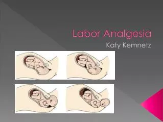
The Physiology of Pain in Labor. 1st stage of labor

E N D
1. Labor Epidural Analgesia Dmitry Portnoy, MD
Anesthesiology Department
2. The Physiology of Pain in Labor 1st stage of labor � mostly visceral
Dilation of the cervix and distention of the lower uterine segment
Dull, aching and poorly localized
Slow conducting, visceral C fibers, enter spinal cord at T10 to L1
2nd stage of labor � mostly somatic
Distention of the pelvic floor, vagina and perineum
Sharp, severe and well localized
Rapidly conducting A-delta fibers, enter spinal cord at S2 to S4
3. The Intensity of Pain in Labor
4. Boundaries of the Epidural Space Superior - the foramen magnum
Inferior limit - the sacral hiatus and sacro-coccygeal membrane
Anterior - the posterior longitudinal ligament covering the bodies of the vertebrae and the intervertebral discs
Posterior - periosteum of laminae of the vertebrae and the ligamenta flava
Lateral � periosteum of the pedicles and intervertebral foraminae
5. Spread of Epidurally Injected Solutions Epidurally administered drugs must travel through:
dura matter arachnoid matter
CSF pia matter
white matter gray matter
Rapid access via �dural cuff�
Competing pathways:
Uptake into epidural epidural fat
Uptake into systemic circulation
6. Indications for LEA PAIN EXPERIENCED BY A WOMAN IN LABOR
When medically beneficial to reduce the stress of labor
ACOG and ASA stated
� in the absence of a medical contraindication, maternal request is a sufficient medical indication for pain relief��
Points of controversy
When?
Who?
How?
7. Contraindications for LEA ABSOLUTE
Patients refusal
Inability to cooperate
Increased intracranial pressure 2 mass lesion
Infection at the site of needle placement
Frank coagulopathy
Severe hypovolemia
Inadequate training RELATIVE
Systemic maternal infection
Preexisting neurological deficiency
Mild or isolated coagulation abnormalities
Relative (and correctable) hypovolemia
Poor communication
8. We are All Ready�Now What? - Last Check! Obstetrician is consulted and confirmed LEA
Preanesthetic evaluation is performed/verified
Pt�s (and only patient�s) desire to have LEA is reconfirmed
Pt�s understanding of risks of LEA is reconfirmed
Fetal well-being is assessed and reassured (obstetrician?, midwife?, yourself?)
Supporting personal is available and present
Resuscitation equipment and drugs are immediately available in the area where LEA placed
9. Standard Technique of LEA Pre epidural check list is completed
Aspiration prophylaxis (?) UTMB � 30 cc Bicitra
Intravenous hydration (What? When? How?)
Monitoring
BP every 1 to 2 min for 20 min after injection of drugs
Continuous maternal HR during induction ( e.g., pulse oximetry)
Continuous FHR monitoring
Continual verbal communication
Maternal position ( sitting or lateral?)
Sterile technique � not negotiable
10. Standard Technique of LEA (cont.) 7. Loss-of-resistance technique of your choice
Catheter is threaded 3 to 5 cm into the space
Secure taping (sponge? tegaderm? loop? tape?)
Testing the catheter
Aspiration test (say NO to big syringe!)
Test dose (what? when? how?)
11. Inducing LEA ( Treat every bolus as a test dose!)
12. Assessment of LEA (sensory, motor, autonomic)
13. Repeat assessment every 1 to 2 hours
12. Etiology and Contributing Factors (Anatomical considerations) Midline epidural structures
plica mediana dorsalis (dura matris) - Luyendijk , 1963, epidurography
midline adhesion of dura mater - Singh, 1967
epidural plica mediana dorsalis - Savolaine, 1988 using CT
dorsomedian connective tissue band - Blomberg, 1986, epiduroscopy
median epidural septum
Connective tissue plane on both dorsolateral compartments of the epidural space - Gallart, 1990
Spinal nerve root diameter - Galindo, 1975
13. Etiology and Contributing Factors (Technique, methodology and equipment) Initial catheter misplacement - incorrect placement
Malposition in anterior or paravertebral (lateral) epidural space
Transforaminal escape
Increased skin-to-epidural space distance
Catheter related
Catheter migration after initial proper placement
The distance of insertion inside the epidural space
Uniport versus multiport epidural catheters
Catheter malfunction and catheter defects
Air for loss-of-resistance technique
Method of injecting local anesthetic
Patient�s position
14. Etiology and Contributing Factors(Patient-related and other risk factors) Inherited and acquired anatomical features
Morbid obesity and body mass index greater than 30
Short and tall individuals
Previous spinal surgery and a variety of musculoskeletal disorders
History of a previous placement of epidural catheter
Radicular pain during epidural placement
Posterior presentation of the fetus
Inadequate analgesia from the initial dose
Duration of labor more than 6 hours
Technical skills, or performance factor
15. Unsatisfactory Labor Epidural AnalgesiaManagement Options Catheter manipulation
Additional volume of local anesthetic
Patient�s position manipulation
Replacement of the epidural catheter
A single shot spinal anesthesia
Continuous spinal anesthesia
Combined spinal-epidural anesthesia
Placement of an additional epidural catheter
Supplementation with intravenous medications
16. Management of Unsatisfactory Epidural
17. Management of Unsatisfactory Epidural
18. Labor Epidural Pearls (Humble Suggestions) Not always epidural is worth its risks
Do not insist unless medically indicated
Consider other pain control options when LEC placement is risky
No epidural analgesia with instant onset (not even close to)
Realistic expectations and labor dynamics
Constant communication during procedure
Treat every dose as a test dose
The longer skin-to-epidural distance, the deeper catheter inside the space
Do not allow the level to recede
19. Avoiding Epidural Disasters (made ridiculously simple) Maintain constant verbal contact with patient
Always aspirate before each injection
Observe for passive return through the catheter
Do not inject more than 4 ml of LA at a time
Observe the patient at least 1.5-2 min between boluses
If in doubts, repeat test dose. Still in doubts? Replace it
After all, be mentally prepare to treat
Convulsions
Total spinal
Cardiovascular collapse and arrest