Download
1 / 51
580 likes | 892 Vues
The Integumentary System. / biology/Wags/ histopage/colorpage/cin/ cin.htmhttp://www.udel.edu. Picture From: http://www.myyogaonline.com/healthy-living/natural-beauty/structure-function-and-care-of-human-skin. The Integumentary System.

E N D
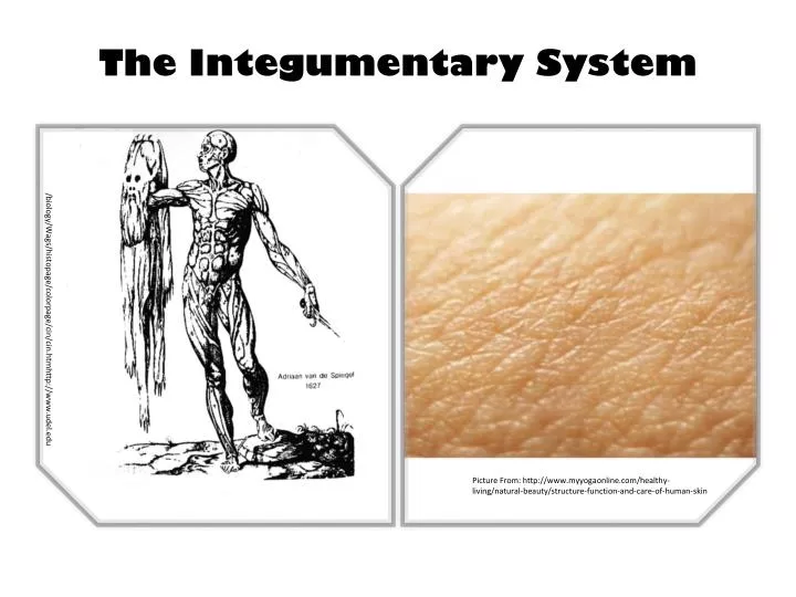
The Integumentary System /biology/Wags/histopage/colorpage/cin/cin.htmhttp://www.udel.edu Picture From: http://www.myyogaonline.com/healthy-living/natural-beauty/structure-function-and-care-of-human-skin
The Integumentary System • composed of the skin, hair, oil and sweat glands, nails, and sensory receptors. • helps maintain a constant body temperature, protects the body, and provides sensory information about the surrounding environment. • Helps to maintain homeostasis • Any change in homeostasis is represented by a change in physical characteristics of the skin
How do we study the skin? • Dermatology (der-ma-TOL-o¯-je¯; dermato- skin; -logy study of ) • Is the medical specialty that deals with the diagnosis and treatment of integumentary system disorders.
Structure of the Skin • The skin, also known as the cutaneous membrane • covers the external surface of the body and is the largest organ of the body in both surface area and weight.
Skin (Integument) • Consists of three major regions • Epidermis – outermost superficial region • Dermis– middle region • Hypodermis– deepest region
Epidermis • Composed of keratinized stratified squamous epithelium, consisting of four distinct cell types and four or five layers • Cell types include keratinocytes, melanocytes, Merkel cells, and Langerhans’ cells • Outer portion of the skin is exposed to the external environment and functions in protection
Cells of the Epidermis • Keratinocytes – produce the fibrous protein keratin • Melanocytes – produce the brown pigment melanin • Langerhans’ cells – epidermal macrophages that help activate the immune system • Merkel cells – function as touch receptors in association with sensory nerve endings
Cells of the Epidermis Keratinocytes – produce the fibrous protein keratin Melanocytes – produce the brown pigment melanin Langerhans’ cells – epidermal macrophages that help activate the immune system Merkel cells – function as touch receptors in association with sensory nerve endings
Layers of the Epidermis: Stratum Basale (Basal Layer) • Deepest epidermal layer firmly attached to the dermis • Consists of a single row of the youngest keratinocytes • Cells undergo rapid division, hence its alternate name, stratum germinativum • Some cells in this layer are stem cells that undergo cell division to continually produce new keratinocytes. • The nuclei of keratinocytes in the stratum basale are large.
Layers of the Epidermis: Stratum Basale (Basal Layer) http://www.imperial.edu/~thomas.morrell/cha_5_tortora_integument.htm
Layers of the Epidermis:Stratum Spinosum (Prickly Layer) • Cells contain a weblike system of intermediate filaments attached to desmosomes • Melanin granules and Langerhans’ cells are abundant in this layer • This stratum mainly consists of numerous keratinocytes arranged in 8–10 layers. • Cells in the more superficial layers become somewhat flattened. • The keratinocytesin the stratum spinosum, which are produced by the stem cells in the basal layer, have the same organelles as cells of the stratum basale and some retain their ability to divide.
Layers of the Epidermis: Stratum Granulosum (Granular Layer) • Thin; three to five cell layers in which drastic changes in keratinocyte appearance occurs • Flattened keratinocytes that are undergoing apoptosis • Keratohyaline and lamellated granules accumulate in the cells of this layer • Keratin – Fibrous protein found in epidermis, hair and nails making them hard and water-repellent. Precursor of keratohyaline.
Layers of the Epidermis: Stratum Lucidum (Clear Layer) • Thin, transparent band superficial to the stratum granulosum • Consists of a few rows of flat, dead keratinocytes • Present only in thick skin • areas such as the fingertips, palms, and soles. Picture from:denisehandlon.hubpages.com
Layers of the Epidermis: Stratum Corneum (Horny Layer) • Outermost layer of keratinized cells • Accounts for three quarters of the epidermal thickness • Functions include: • Waterproofing • Protection from abrasion and penetration • Rendering the body relatively insensitive to biological, chemical, and physical assaults
KeratinizationGrowth of the Epidermis • Newly formed cells in the stratum basale are slowly pushed to the surface. • As the cells move from one epidermal layer to the next, they accumulate more and more keratin, a process called keratinization. • The mechanisms that regulate this remarkable growth are not well understood, but hormonelike proteins such as epidermal growth factor (EGF) play a role. • An excessive amount of keratinized cells shed from the skin of the scalp is called dandruff.
Dermis • Second major skin region containing strong, flexible connective tissue • Cell types include fibroblasts, macrophages, and occasionally mast cells and white blood cells • Composed of two layers: • Papillary • Reticular
Layers of the Dermis • Papillary layer • Areolar connective tissue with collagen and elastic fibers • Its superior surface contains peglike projections called dermal papillae • Dermal papillae contain capillary loops, Meissner’s corpuscles, and free nerve endings
Layers of the Dermis • Reticular layer • Accounts for approximately 80% of the thickness of the skin • Collagen fibers in this layer add strength and resiliency to the skin • Elastin fibers provide stretch-recoil properties
Hypodermis • Subcutaneous layer deep to the skin • Composed of adipose and areolar connective tissue
Cellulite http://hubpages.com/hub/science_of_cellulite http://glamouredited.com/skin/tips_for_removing_cellulite.html http://www.theperfectphit.com/images/velashape2.jpg
Fingerprint Sweat Glands
Sebaceous Glands • Simple alveolar glands found all over the body • Soften skin when stimulated by hormones • Secrete an oily secretion called sebum
Skin Color • Three pigments contribute to skin color • Melanin – yellow to reddish-brown to black pigment, responsible for dark skin colors • Freckles and pigmented moles – result from local accumulations of melanin • Carotene – yellow to orange pigment, most obvious in the palms and soles of the feet • Hemoglobin – reddish pigment responsible for the pinkish hue of the skin
Hair • Filamentous strands of dead keratinized cells produced by hair follicles • Contains hard keratin which is tougher and more durable than soft keratin of the skin • Made up of the shaft projecting from the skin, and the root embedded in the skin • Consists of a core called the medulla, a cortex, and an outermost cuticle • Pigmented by melanocytes at the base of the hair
Hair Function and Distribution • Functions of hair include: • Helping to maintain warmth • Alerting the body to presence of insects on the skin • Guarding the scalp against physical trauma, heat loss, and sunlight • Hair is distributed over the entire skin surface except • Palms, soles, and lips • Nipples and portions of the external genitalia
Types of Hair • Vellus– pale, fine body hair found in children and the adult female • Terminal – coarse, long hair of eyebrows, scalp, axillary, and pubic regions
Sweat Glands • Different types prevent overheating of the body; secrete cerumen and milk • Eccrine (merocrine) sweat glands – found in palms, soles of the feet, and forehead • Apocrine sweat glands – found in axillary and anogenital areas • Ceruminous glands – modified apocrine glands in external ear canal that secrete cerumen • Mammary glands – specialized sweat glands that secrete milk
Structure of a Nail • Scalelike modification of the epidermis on the distal, dorsal surface of fingers and toes Figure 5.4
Functions of the Integumentary System • Protection – chemical, physical, and mechanical barrier • Body temperature regulation is accomplished by: • Dilation (cooling) and constriction (warming) of dermal vessels • Increasing sweat gland secretions to cool the body • Cutaneous sensation – exoreceptors sense touch and pain
Functions of the Integumentary System • Metabolic functions – synthesis of vitamin D in dermal blood vessels • Blood reservoir – skin blood vessels store up to 5% of the body’s blood volume • Excretion – limited amounts of nitrogenous wastes are eliminated from the body in sweat
DEVELOPMENT OF THE INTEGUMENTARY SYSTEM
Burns • First-degree – only the epidermis is damaged • Symptoms include localized redness, swelling, and pain • Second-degree – epidermis and upper regions of dermis are damaged • Symptoms mimic first degree burns, but blisters also appear • Third-degree – entire thickness of the skin is damaged • Burned area appears gray-white, cherry red, or black; there is no initial edema or pain (since nerve endings are destroyed)
Rule of Nines • Estimates the severity of burns • Burns considered critical if: • Over 25% of the body has second-degree burns • Over 10% of the body has third-degree burns • There are third-degree burns on face, hands, or feet