Download
1 / 20
350 likes | 1.47k Vues
Phenylketonuria (PKU). Sanketh Proddutur. What is PKU?. Phenylketonuria (PKU) is an inherited error of metabolism caused by a deficiency in the enzyme phenylalanine hydroxylase (PAH). phenylalanine hydroxylase converts the amino acid phenylalanine to tyrosine, another amino acid . . Pathology.

E N D
Phenylketonuria (PKU) Sanketh Proddutur
What is PKU? • Phenylketonuria (PKU) is an inherited error of metabolism caused by a deficiency in the enzyme phenylalanine hydroxylase (PAH). • phenylalanine hydroxylase converts the amino acid phenylalanine to tyrosine, another amino acid.
Pathology • mutations in the human gene (chromosome 12.q22-24.2), encoding PAH result in the autosomal recessively inherited disease hyperphenylalaninemia (HPA) • The resulting phenotypes can range in severity from mild hyperphenylalaninemia (HPA) • classic PKU which inevitably leads to mental retardation if left untreated.
Symptoms Newborn infants develop signs of PKU within a few months of birth if untreated: • Mental retardation • Behavioral or social problems • Seizures, tremors or jerking movements in the arms and legs • Rocking • Hyperactivity • Stunted growth
Symptoms (contnd) • Skin rashes (eczema) • Small head size (microcephaly) • Vomiting • A musty odor in the child's breath, skin or urine • Fair skin and blue eyes
Etiology Human phenylalanine hydroxylase converts the essential amino acid L-phenylalanine (L-Phe) into L-tyrosine using the co-factor (6R)-L-erythro-5,6,7,8-tetrahydrobiopterin (BH4) and molecular oxygen. Any disruption in this mechanism can lead to Hyperphenylalaninemia.
Etiology (contnd) 99% of the mutations that disrupt this mechanism can be traced back to the PAH gene, 1% of the mutations are in the genes encoding the enzymes which are involved in the regeneration of the cofactor BH4, which is a vital co-substrate of PAH.
BH4 regeneration BH4 is a cofactor of PAH and its regeneration is vital for the ongoing reaction.
PAH Liver PAH is reported to exist in solution as a pH dependent equilibrium between homo-dimers and the active homo-tetramers. Four identical molecules of phenylalanine hydroxylase interact to form the tetramer, which is the functional unit for this enzyme.
PAH (contnd) Each monomer polypeptide is composed of three domains • An N-terminal regulatory domain (residues 1-142) • A central catalytic domain (residues 143-410) which includes the active site iron • A C-terminal tetramerization domain (residues 411-452).
The regulatory domain consists of a four stranded anti-parallel β-sheet flanked on side by two short α-helices and on the other side by the catalytic domain The catalytic domain houses the active site iron in a novel basket like arrangement of 14 α-helices and 8-β strands the short tetramerization domain is formed by a C-terminal "arm" of two -strands forming a -ribbon, and a long -helix and is responsible for assembling the four chains into the tetramer.
Catalytic domain The active site iron is ligated in an octahedral manner and bound to His285, His290, and 1 oxygen atom in Glu330 as well as three water molecules Fe(II) ion His 290 Glu 330 His 285
Catalytic domain (contnd) A tunnel adjacent to the active site might be responsible for directing the substrate.
Catalytic mechanism In order to hydroxylate the unactivated Phenylalanine to Tyrosine, PAH incorporates one atom of oxygen from molecular oxygen into the substrate and reduces the other atom to water. In the process, the BH4 cofactor undergoes a two-electron oxidation to form quinonoid dihydropterin (q-BH2) which is then regenerated back to BH4.
How exactly does this take place? We don’t know! It is however speculated that the binding of the substrate is cooperative, that is the tetramer undergoes synergistic conformational changes to bind the substrates subsequently hydroxylating the Phe residue.
Binding of the cofactor The crystal structure of the dimeric catalytic domain (residues 118-424) of human PAH cocrystallized with the oxidized form of the cofactor (7,8-dihydro-L-biopterin, BH2), has been determined at 2.0 Å resolution. The pterin binds close to the catalytic iron and forms an extensive H-bond network with Ala322, Gly247, Leu249 and Glu286. The phenyl ring of Tyr325 establishes hydrophobic contacts with the pterin, and thus contributes to the correct positioning of the pterin cofactor for catalysis, furthermore this also ensures that the pterin ring forms an aromatic pi-stacking interaction with Phe254.
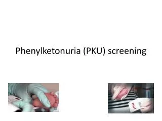
Tests • Guthrie card bloodspot: is the standard test for PKU. Blood taken from the infant’s heel after first breast feeding is assayed for high levels of Phe. • tandem mass spectrometry (often abbreviated as MS/MS). This technology can detect the blood components that are elevated in certain disorders, and is capable of screening for more than 20 inherited metabolic disorders with a single test. • Amniocentesis: is a form of prenatal diagnosis in which DNA extracted from fetal cells obtained from the amniotic fluid at about 15-18 weeks' gestation period.
Treatment • Restriction of dietary phenylalanine: is the most obvious treatment for this disorder and also the most prevalent. Phe is ubiquitous across a variety of food groups but is especially prevalent in proteins. A typical Phe-restricted diet would be rich in fruits and some amounts of potato and milk to maintain a phe concentration of about 2-6 mg/dL supplemented by a phe-free protein supplement.All kinds of meat, cheese and flour products are forbidden. Prognosis: is fairly good if the diet is strictly followed into adulthood. Patients who discontinued after late adolescence. • Other potential treatments being investigated are gene therapy, BH4 and phenylalanine lyase administration.
References • Erlandsen, H. and Stevens, R.C. (1999) The structural basis of phenylketonuria. Molecular Genetics and Metabolism 68, 103-25. • F.Fusetti, H.Erlandsen, T.Flatmark, R.C.Stevens. (1998) Structure of Tetrameric Human Phenylalanine Hydroxylase and its Implications for Phenylketonuria. J.Biol.Chem. 273, 16962 • O.A.Andersen, T.Flatmark, E.Hough. (2001) High Resolution Crystal Structures of the Catalytic Domain of Human Phenylalanine Hydroxylase in its Catalytically Active Fe(II) Form and Binary Complex with Tetrahydrobiopterin. J.Mol.Biol. 314; 266 • H.Erlandsen, E.Bjorgo, T.Flatmark, R.C.Stevens(2000). Crystal Structure and Site-Specific Mutagenesis of Pterin-Bound Human Phenylalanine Hydroxylase. Biochemistry 2208;